How Does Calcium Deficiency Affect Bone Health?

To keep our bones healthy and strong, it’s important to eat a balanced diet with enough nutrients, especially calcium and vitamin D. Calcium is crucial for building and maintaining strong bones and teeth, as well as supporting other body functions like blood circulation and muscle control. Vitamin D is needed to effectively absorb calcium from the food we eat.

Since our bodies can’t produce calcium on their own, it’s essential to get it from the foods we eat. If we don’t consume enough calcium, our bodies will take it from the stored supply in our bones. Over time, this can lead to weakened bones and an increased risk of conditions like osteoporosis, where bones become brittle. Other conditions like osteopenia and hypocalcemia can also result.

In children, insufficient calcium intake may affect their growth potential, preventing them from reaching their full height. Therefore, it’s crucial to ensure that we consume the recommended daily amount of calcium through various food sources, vitamins, and supplements to keep our bones healthy and prevent these issues.

Book your Advance Bone Health test today!

What is Calcium Deficiency?

Calcium deficiency, also known as hypocalcemia, is a condition where the level of calcium in the blood is too low. This can result in issues like osteoporosis, changes in dental health, brain alterations, and the development of cataracts.

How Much Calcium Do You Need Each Day?

Your age determines the appropriate daily amount of calcium you should consume. Here’s what’s suggested for adults:

  • For individuals aged 19 to 50 years, the recommended daily calcium intake is 1000 mg.
  • Men between the ages of 51 and 70 are advised to consume 1000 mg of calcium per day.
  • For women aged 51 to 70 years, the recommended daily calcium intake is 1000 mg.
  • For adults aged 71 years and above, the recommended daily calcium intake is 1200 mg.
  • Pregnant and lactating adults should aim for a daily calcium intake of 1000 mg.

Why is Calcium Important?

Calcium is like a superhero for our bones. About 99% of it hangs out in our bones and teeth, making them strong and hard. The remaining bit of calcium does some other cool stuff in our bodies to keep things running smoothly. It helps blood vessels do their expanding and contracting dance, muscles contract, and the nervous system transmit messages.

Every day, calcium plays a game of musical chairs in our bones, coming in and out as they undergo constant remodeling. In kids and teens, the body adds more bone than it breaks down, making bone mass grow. This party lasts until around age 30 when new bone creation and old bone breakdown balance out. But as we get older, especially after menopause in women, bones break down faster than they’re created. If we don’t get enough calcium in our diet, it can lead to osteoporosis, where bones become fragile and prone to breaking. So, calcium is not just for building strong bones; it’s also a key player in keeping our body’s daily operations in check.

What Leads to Calcium Deficiency?

There are various reasons why someone might have a lack of calcium, such as:

  • Not getting enough calcium in your diet for a while.
  • Having trouble with foods rich in calcium due to dietary intolerance.
  • Genetic factors can play a role.
  • Taking certain medications that reduce calcium absorption.
  • Hormonal changes, especially in postmenopausal women or those who’ve had hysterectomy and oophorectomy.

Signs of Not Enough Calcium in Your Body

When your body lacks enough calcium, it can lead to various effects. At first, there might not be any noticeable symptoms. However, over time, a calcium deficiency can result in low bone density, increasing the risk of brittle bones known as osteoporosis. Osteoporosis is often referred to as a “silent disease” because symptoms may not appear until a bone is broken.

In more advanced stages of osteoporosis, individuals may experience back pain due to fractured or collapsed vertebrae, a gradual loss of height, a stooped posture, and bones that break more easily than expected. Diagnosis typically involves a DEXA bone mineral density test before symptoms become evident.

In cases of acute calcium deficiency, the symptoms can be more severe and include:

  • Muscle spasms
  • Muscle cramps
  • Bones breaking easily
  • Memory loss
  • Confusion
  • Numbness or tingling sensation in the feet, hands, and face
  • Hallucinations
  • Brittle and weak nails
  • Depression

If you notice any of these symptoms, it’s essential to consult with a healthcare professional for proper evaluation and guidance.

Easy Tips for Stronger Bones and Increased Calcium Intake

Boosting your bone health and getting enough calcium doesn’t have to be complicated. Follow these straightforward tips:

  • Meet the Daily Calcium Requirement: Start by meeting the recommended dietary allowance (RDA) of calcium every day. Include calcium-rich foods in your diet, such as soy products (tofu, soy milk, soy drink), dairy products (yogurt, cheese, paneer), vegetables (beans, legumes, broccoli, cabbage, carrot, cauliflower, celery, okra, peas, sweet potato), fruits (berries, dates, figs, orange, papaya), cereals (corn flakes), nuts (almonds, sesame seeds), eggs, and fish.
  • Consider Supplements: If it’s challenging to get enough calcium from your diet alone, consult your doctor about taking supplements.
  • Ensure Balanced Nutrition: It’s not just about calcium; your bones benefit from other nutrients too. Make sure you get enough protein, magnesium, vitamin D, vitamin K2, and phosphorus.
  • Magnesium Matters: Magnesium aids in the absorption and retention of calcium, contributing to stronger bones and helping prevent osteoporosis.
  • Vitamin D Support: Vitamin D plays a crucial role in calcium absorption and regulates its levels in the blood. Make sure you get enough sunlight exposure or consider supplements if needed.
  • Phosphorus Partnership: Phosphorus, working alongside calcium, also plays a role in maintaining bone health. Don’t overlook its importance.
  • Consult a Dietician: For a personalized approach, consult with a nutrition expert. They can help you create a well-balanced diet that includes the right amounts of calcium, phosphorus, and vitamin D.

By incorporating these tips into your routine, you can promote strong bones and overall bone health.

How Smoking Hurts Bones and Osteoporosis

When you smoke, it makes it harder for your body to soak up calcium, which results in less bone density and weaker bones. The nicotine in cigarettes slows down the creation of the cells responsible for building strong bones, making it tougher for your body to heal.

Can Drinking Too Much Alcohol Decrease Vitamin D?

If you regularly drink a lot of alcohol, it can mess with how your body handles vitamin D. People who are chronic alcoholics often end up with low levels of a type of vitamin D called 25-hydroxyvitamin D (25(OH)D). Also, it’s worth noting that alcohol can up the chances of falls in older adults dealing with osteoporosis, which can result in serious fractures—the most severe outcome of this bone condition.


Coronary Artery Disease

Coronary artery disease (CAD) is a critical health condition that impacts the blood vessels responsible for supplying oxygen-rich blood to the heart. Recognizing the risk factors associated with CAD is crucial for early detection and prevention. Factors such as high blood pressure, elevated cholesterol levels, smoking, obesity, diabetes, family history of heart disease, and a sedentary lifestyle can significantly increase the likelihood of developing coronary artery disease.

Consulting with the best cardiologist in Delhi is paramount when it comes to understanding the risk factors and symptoms of CAD. Ayushman Hospital, renowned for its team of highly skilled and experienced heart specialists, stands as a beacon for providing exceptional care and guidance. If you suspect any symptoms, seeking consultation with a cardiologist in Delhi is crucial for early intervention.

Coronary Artery Disease Symptoms

Recognizing the symptoms of CAD is equally essential for timely intervention. The symptoms include:

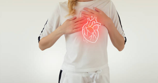
  • Chest pain or discomfort (angina)
  • Shortness of breath
  • Fatigue
  • Dizziness
  • Nausea
  • Sweating
  • Palpitation

These common signs should never be ignored. Seeking consultation with a cardiologist at Ayushman Hospital, known for its expertise in cardiovascular care, ensures an accurate diagnosis and personalized treatment plans tailored to individual needs. Trusting the best heart specialist in Delhi is paramount for comprehensive care and support.

Lifestyle Changes for Coronary Artery Disease Prevention

Adapting certain lifestyle changes can significantly contribute to the prevention of CAD. The following modifications are recommended:

  • Don’t Smoke: Smoking remains one of the leading risk factors for coronary artery disease. Quitting smoking is the most beneficial action for heart health.
  • Eat a Heart-Healthy Diet: A heart-healthy diet involves reducing the intake of saturated and trans fats, cholesterol, and sodium while increasing the consumption of fruits, vegetables, and whole grains.
  • Be Physically Active: Strive for at least 150 minutes of moderate-intensity aerobic activity or 75 minutes of vigorous-intensity aerobic activity weekly. Incorporating shorter sessions throughout the day is also effective.
  • Maintain a Healthy Weight: Being overweight or obese increases the risk of CAD. Even a small amount of weight loss can significantly improve heart health.
  • Manage Stress: Stress is linked to increased blood pressure and cholesterol levels, elevating the risk of CAD. Adopt healthy stress management techniques such as exercise, relaxation, and spending time with loved ones.
  • Regular Check-Ups: Regular medical check-ups are crucial for understanding and monitoring heart health, and preventing lifestyle disorders like CAD. Consult with a cardiologist in Delhi for personalized advice on maintaining heart health.
  • Consultation and Online Services: Taking the first step towards proper care involves consulting an expert. For any health concerns, it is recommended to undergo an examination by a certified medical professional or visit the finest cardiology hospital in Delhi. Additionally, online consultations with experts at Ayushman Hospital are available for expert advice and guidance.

Conclusion:

Coronary artery disease is a serious health condition that demands attention and proactive measures. Understanding the risk factors, recognizing symptoms, and adopting a heart-healthy lifestyle are key components of prevention. Trusting the expertise of the best cardiologist in Delhi ensures accurate diagnosis and personalized care.

By implementing lifestyle changes, such as quitting smoking, adopting a heart-healthy diet, staying physically active, maintaining a healthy weight, managing stress, and prioritizing regular check-ups, individuals can significantly reduce their risk of coronary artery disease. Taking preventive actions and seeking guidance from the finest cardiology hospital in Delhi are crucial steps towards a heart-healthy life. If you suspect any symptoms, consult with a cardiologist in Delhi for personalized advice and timely intervention.

Tuberculosis

Tuberculosis, commonly known as TB, remains a longstanding and formidable health challenge that has captured the attention of medical professionals and researchers throughout history. In India, approximately 40% of the population is affected by TB bacteria, with the majority harboring latent rather than active TB. Shockingly, the World Health Organization (WHO) estimates that 300,000 people succumb to TB annually in India. According to the German Leprosy & Tuberculosis Relief Association (DAHW), one-fifth of the world’s new tuberculosis cases are concentrated in the Indian subcontinent.

Recognizing the severity of the situation, Tuberculosis or TB emerges as one of India’s most significant health concerns today. Every citizen must be well-informed about this ailment. This article delves into various facets of tuberculosis, aiming to provide a comprehensive understanding of the disease and highlight the progress made in its prevention and treatment.

What is Tuberculosis

Tuberculosis is a bacterial infection primarily targeting the lungs and potentially spreading to various organs, including the kidneys, spine, and brain. Without proper treatment, TB disease can prove fatal. While historically linked to the impoverished, today, it is recognized as affecting individuals across all social classes.

What Leads to Tuberculosis?

Tuberculosis is the microorganism responsible for the onset of tuberculosis. This germ can stay in the air for a long time. That’s why people with tuberculosis need to be careful and take steps to stop the germ from spreading to others.

How Tuberculosis Spreads

Tuberculosis mostly spreads through the air. When someone with active TB disease coughs or talks, tiny drops from their breath, carrying the bacteria Mycobacterium tuberculosis, can float in the air. The main ways TB can be passed on include:

How Tuberculosis Spreads Easily Explained

  • Airborne Transmission: Tuberculosis mainly spreads when someone with active TB coughs, sneezes, talks, or sings, releasing tiny droplets containing the infectious bacteria into the air. Inhaling these droplets can lead to infection with Mycobacterium tuberculosis.
  • Close and Prolonged Contact: TB is more likely to spread when people spend a lot of time near an infectious person. This is especially true in crowded or enclosed places like homes, prisons, or healthcare facilities, where there’s a higher concentration of these respiratory droplets in the air.
  • Poor Ventilation: In places with inadequate airflow, infectious droplets can linger in the air, increasing the risk of TB transmission. Good ventilation helps disperse and dilute these droplets, lowering the chances of TB spreading.
  • Weakened Immune System: People with weakened immune systems, like those with HIV/AIDS, malnutrition, or undergoing immune-suppressing treatments, are more vulnerable to TB infection. They have a higher risk of progressing from latent TB infection to active TB disease.

It’s crucial to know that not everyone exposed to Mycobacterium tuberculosis gets sick. Some may have latent TB infection, where the bacteria are present but inactive, causing no symptoms and not spreading to others. However, if their immune system weakens, the latent infection can turn into active TB disease, which is contagious.

Types of Tuberculosis

Tuberculosis (TB) comes in different forms, mainly divided into latent TB infection (LTBI) and active TB disease.

Latent TB Infection (LTBI): In this type, people have the TB bacteria, but it’s inactive. They don’t show symptoms and can’t spread it to others. However, there’s a risk it could turn into active TB disease, especially in people with weakened immune systems.

Active TB Disease: This occurs when the bacteria become active, causing symptoms. It can affect the lungs (pulmonary TB) or other body parts (extrapulmonary TB). Symptoms include cough, chest pain, weight loss, fatigue, fever, and night sweats. TB in its active form is infectious and can be transmitted through the air.

Additional TB Categories: 

  • Pulmonary TB: Affects the lungs, causing cough, chest pain, and breathing issues.
  • Extrapulmonary TB: Affects parts outside the lungs, like kidneys, spine, lymph nodes, and brain.
  • Miliary TB: A severe form where bacteria spread through the bloodstream to multiple organs.
  • Drug-resistant TB: Some TB strains resist common antibiotics, making treatment more challenging.

Understanding these types is crucial for diagnosing, treating, and preventing tuberculosis effectively.

Recognizing Signs and Symptoms of Tuberculosis

Tuberculosis can show various signs and symptoms, depending on whether it’s latent or active. Here are common indicators linked to active TB disease:

  • Chest Pain: Feeling pain in the chest, especially during deep breaths or coughing, could be due to lung tissue inflammation.
  • Persistent Cough: A cough lasting three weeks or more is typical for pulmonary TB. It may produce sputum (mucus), possibly with blood.
  • Unexplained Weight Loss: Active TB often leads to weight loss due to factors like reduced appetite and increased metabolic demands during infection.
  • Fatigue: Persistent fatigue and weakness are common as the body fights the infection.
  • Fever: A low-grade or persistent fever, accompanied by chills, especially in the afternoon or evening, can signal active TB.
  • Night Sweats: Profuse sweating, especially at night, is a characteristic TB symptom that can disrupt sleep.
  • Shortness of Breath: Difficulty breathing may occur, especially if the infection has progressed and affected a significant part of the lungs.
  • Swollen Lymph Nodes: In extrapulmonary TB, where organs outside the lungs are involved, swollen lymph nodes may be noticeable in various body parts.

If someone shows these symptoms or has been in close contact with someone diagnosed with active TB, seeking prompt medical attention is crucial for proper evaluation, diagnosis, and treatment. Early detection and treatment are vital for effectively managing TB and preventing its spread to others.

How is Tuberculosis Diagnosed?

Diagnosing tuberculosis involves a combination of clinical assessment, imaging scans, and laboratory tests. Here are the common tests used to diagnose TB:

  • Tuberculin Skin Test (TST) or Mantoux Test: A small amount of purified protein is injected under the skin. If a raised bump appears, it indicates exposure to TB, but it can’t differentiate between latent and active TB.
  • Interferon-gamma Release Assays (IGRAs): Blood tests like QuantiFERON-TB Gold or T-SPOT.TB measures the release of interferon-gamma in response to TB-specific antigens. They are more specific than the skin test and help distinguish between latent and active TB.
  • Chest X-ray: Imaging the chest helps identify lung abnormalities associated with pulmonary TB, such as infiltrates or cavities.
  • Sputum Smear Microscopy: Examining mucus samples under a microscope helps detect the presence of TB bacilli.
  • Sputum Culture: Culturing sputum samples confirms TB and enables drug susceptibility testing.
  • Nucleic Acid Amplification Tests (NAATs): Molecular tests like PCR detect TB genetic material in clinical samples, providing a rapid and sensitive diagnosis.
  • Xpert MTB/RIF Assay: This molecular test simultaneously detects TB and assesses rifampicin resistance, offering quick results.
  • Bronchoscopy: When sputum samples are hard to obtain, bronchoscopy collects samples from the lower respiratory tract.
  • Biopsy: For extrapulmonary TB, a biopsy of affected tissues or organs may be performed for microscopic examination and culture.
  • Drug Susceptibility Testing (DST): This test determines TB bacteria’s susceptibility to specific antibiotics and guiding treatment.

The choice of tests depends on factors like clinical presentation, risk factors, and available resources. Multiple tests may be used for accurate diagnosis, especially for drug-resistant strains or extrapulmonary TB.

How to Prevent Tuberculosis?

Preventing tuberculosis involves a combination of strategies to reduce transmission, detect and treat latent and active infections, and address risk factors. Here are key measures for TB prevention:

  • BCG vaccine: Administer the Bacillus Calmette-Guérin (BCG) vaccine in many countries to reduce the risk of severe TB complications in children, even though it may not offer complete protection.
  • Respiratory hygiene: Encourage good respiratory hygiene, like covering the mouth and nose when coughing or sneezing. Use masks in crowded public places, such as trains, buses, and stations.
  • Ventilation: Ensure well-ventilated spaces, particularly in crowded areas, to decrease the concentration of infectious droplets. Use fans or open windows for cross-ventilation.
  • Identifying and treating latent TB infection: Screen individuals at higher risk for TB, including those with known exposure or immunosuppression. Offer to test for latent TB infection (LTBI) and provide appropriate treatment to prevent progression to active TB.
  • Contact tracing and testing: Identify and test individuals in close contact with someone diagnosed with active TB. This aids in the early detection and treatment of latent or active TB in those exposed.
  • Public health education: Increase public awareness about TB transmission, symptoms, and the importance of seeking medical care. Educate communities about available testing and treatment services.

Despite being highly contagious, tuberculosis can be prevented by taking these simple precautions in the long run.

Conclusion

Tuberculosis is a significant health challenge that requires everyone’s efforts in prevention, early detection, and effective treatment. If you or someone you know shows symptoms of tuberculosis, it’s crucial to seek medical help promptly. Ayushman Hospital, known for its commitment to excellent healthcare, offers specialized services for diagnosing and treating tuberculosis. Our experienced specialists use advanced diagnostics and proven treatments to provide thorough care, ensuring the best outcomes for patients. Remember, taking timely action can make a big difference in managing and controlling tuberculosis. Consult a tuberculosis specialist at Ayushman Hospitals for expert, personalized, and compassionate care.

Neurology Hospital in Delhi

Facing neurological disorders can be overwhelming, and finding the right specialist is crucial for optimal care. Fortunately, we boast renowned medical advancements, offering a wealth of top-notch neurology hospital in Delhi. This guide empowers you to navigate this process effectively, ensuring informed decisions regarding your neurological healthcare needs.

Research About Neurology Hospital in Delhi

Conducting thorough research is imperative when exploring neurology hospitals for your health needs. Various channels can provide valuable insights into the reputation and quality of such healthcare facilities in Delhi.

Online Hospital Reviews: Leverage dedicated online platforms that review hospitals. These platforms allow individuals and their families to share their experiences and rate different hospitals. Pay attention to overall ratings, patient feedback, and specific comments on neurology departments and specialists.

Seek Recommendations from Trusted Sources: Reach out to your primary care physician or other reliable healthcare professionals for recommendations. They can provide valuable perspectives derived from their knowledge and experiences at a neurology hospital in Delhi. Furthermore, seek advice from friends, family members, or colleagues who have undergone neurology treatments in the city. Personal recommendations provide firsthand information about the level of care and patient experience.

Consider Hospital Accreditations: Accreditations and certifications from reputable organizations serve as indicators of a hospital’s commitment to quality and patient safety. Look for neurology hospital in Delhi that have received accreditations from recognized bodies such as the Joint Commission International (JCI) or the National Accreditation Board for Hospitals & Healthcare Providers (NABH). These accreditations signify that the hospital adheres to stringent standards in terms of infrastructure, medical protocols, and patient care.

Choosing the Right Hospital for Your Neurological Care in Delhi

Selecting the best neurology hospital in Delhi is crucial for receiving optimal care for your specific needs. Here are some key factors to consider when making this important decision:

  1. Experienced and Specialized Neurologists: Look for hospitals with a team of highly-skilled neurologists who specialize in various neurological disorders. This ensures you have access to experts who can effectively address your specific needs, whether it’s stroke management, movement disorders, pediatric neurology, or any other area.
  2. Advanced Diagnostic Capabilities: Accurate diagnosis forms the foundation of successful treatment. Ensure the hospital utilizes state-of-the-art diagnostic technology, such as high-resolution MRI and CT scan machines. These advanced tools enable detailed evaluations, allowing neurologists to create precise treatment plans tailored to each patient’s condition.
  3. Dedicated Departments for Specialized Care: neurological disorders present diversely, often requiring specialized care. Choose a hospital with dedicated departments catering to specific neurological disorders, such as epilepsy treatment, neuro-oncology, or neuromuscular disorders. This ensures access to the expertise and resources necessary for comprehensive and effective treatment.
  4. Patient Reviews and Testimonials: While not the sole factor, patient experiences can offer valuable insights. Consider reading testimonials from individuals who have received care at the hospital. Positive feedback can highlight the expertise of the neurology team, the effectiveness of treatments, and the overall patient experience, including factors like compassion and communication.

Remember, thorough research and a clear understanding of your specific needs empower you to make informed decisions for your neurological healthcare journey in Delhi.

Ayushman Hospital: Offering Comprehensive Neurological Care

Ayushman Hospital provides comprehensive neurological care for a wide range of conditions, including epilepsy, neuro-oncology, and neuromuscular disorders.

Key offerings include: 

  • A team of skilled neurologists: The hospital employs experienced neurologists specializing in various neurological areas, ensuring patients receive targeted expert care.
  • Cutting-edge diagnostics: The hospital utilizes advanced technologies like high-resolution MRI and CT scans for accurate diagnoses, forming the foundation for effective treatment plans.
  • Specialized departments: Dedicated departments cater to specific neurological disorders, providing patients with focused expertise and resources for optimal outcomes.

By offering these services, Ayushman Hospital aims to provide patients with a complete and effective neurological care experience.

robotic knee surgery

Once enjoyed dancing, hiking, and taking stairs with ease, but now struggling due to persistent knee pain. Robotic knee surgery is a groundbreaking advancement in orthopedics, providing precise and efficient treatment. This technology, combining robotics with surgical expertise, offers faster recovery, enhanced accuracy, and improved long-term outcomes, restoring the freedom to move and enjoy life.

What is Robotic Knee Replacement Surgery?

Both robotic knee replacement and traditional knee replacement involve an orthopedic surgeon removing damaged knee tissue and replacing it with an artificial joint. The distinction lies in the use of a robotic arm for assistance in robotic knee surgery. This advanced technique provides superior soft tissue balance and joint alignment. If you qualify for robotic surgery, your surgeon will thoroughly discuss all knee replacement options, offering benefits such as improved soft tissue balance, better joint alignment, and various other advantages.

  • Shorter hospital stays.
  • Faster recovery times.
  • Improved range of motion.
  • Higher patient satisfaction.
  • Reduced risk of complications.

How Robotic Knee Surgery Works?

Robotic knee surgery involves the use of a sophisticated robotic system to assist surgeons during the procedure. The knee replacement process typically starts with a detailed preoperative planning phase. Advanced imaging like CT scans is utilized to create a three-dimensional model of the patient’s knee. This model allows your surgeon to meticulously plan the surgery, taking into account the unique anatomy and pathology of the individual.

During the surgery, the robotic system acts as an extension of the surgeon’s skills. It provides real-time data and assists in executing the pre-planned surgical steps with unparalleled precision. The robotic arm is equipped with a range of instruments that can be controlled by your surgeon.

Best Candidate For Robotic Knee Replacement

Not every patient is an ideal candidate for robotic knee surgery. People with osteoarthritis or other degenerative knee conditions can be suitable for this advanced procedure. As this innovative technology continues to gain prominence, it’s important to understand who is the best candidate for robotic knee replacement.

Severity of Knee Damage: Candidates with severe knee damage due to rheumatoid arthritis, osteoarthritis, or traumatic injuries robotic knee replacement is the best option for them. The technology allows for meticulous planning and execution, ensuring optimal outcomes even in complex cases.

Overall Health and Fitness: Good overall health is important for a successful recovery. Candidates with well-managed medical conditions like diabetes or cardiovascular issues, are generally better suited for the procedure. Moreover, people who are non-smokers and maintain a healthy body weight tend to have fewer complications.

Preoperative Range of Motion: Candidates with a reasonably good preoperative range of motion in the knee joint may achieve better results. Robotic knee replacement can enhance joint mechanics and functionality, but starting with a better baseline often leads to more favorable outcomes.

Patient Commitment to Rehabilitation: Commitment to postoperative rehabilitation is a key determinant of success. Candidates who are dedicated to following their rehabilitation protocols, including physical therapy and exercise regimens are more likely to regain strength and mobility faster.

Customization Needs: Patients with unique anatomical considerations, like deformities or variations, benefit greatly from the customization capabilities of robotic surgery. The precision of the technology allows for tailored solutions, ensuring a better fit and alignment of the artificial joint.

Previous Surgical History: People with a limited history of knee surgeries may find robotic knee replacement particularly advantageous. The technology’s ability to adapt to individual anatomies and minimize invasiveness can be especially beneficial in cases where preserving existing structures is essential.

Difference Between Traditional and Robotic Knee Surgery

Here’s a simplified table highlighting the key differences between robotic and traditional knee replacement surgery:

Feature Robotic Knee Replacement Traditional Knee Replacement
Procedure Assisted by robotic technology Manual execution by the surgeon
Preoperative Planning Precise 3D mapping of the joint Standard X-rays and measurements
Incision Size Often smaller incisions Larger incisions
Accuracy Enhanced precision with robotics Manual precision
Bone Resection More precise bone removal Manual bone resection
Implant Placement Guided by the robotic system Surgeon’s experience and skill
Real-time Feedback Continuous feedback during surgery Limited real-time information
Recovery Time Potentially shorter recovery Standard recovery period
Postoperative Pain May experience less pain Typical post-surgery discomfort
Knee Surgery Cost Generally higher costs Typically lower costs

It’s crucial to note that the choice between traditional and robotic knee replacement depends on various factors. The factors include the patient’s condition, the surgeon’s expertise, and available technology. You should always consult with a healthcare professional to determine the most suitable surgery for your case.

Advantages of Robotic Knee Surgery

Studies have shown that robotic knee surgery offers several advantages over traditional surgery, including:

  • Precision and Accuracy: One of the primary advantages of robotic knee surgery is its unmatched precision. The robotic system enables surgeons to make highly accurate cuts and placements.
  • Customization: Each patient’s knee is unique and robotic surgery allows for a high degree of customization. The preoperative planning based on detailed imaging, ensures that the surgery is tailored to the individual’s anatomy and specific pathology.
  • Minimally Invasive: Robotic knee surgery often involves smaller incisions compared to traditional methods. This can result in reduced trauma to surrounding tissues, leading to faster recovery times and less postoperative pain.
  • Faster Recovery: The precision and minimal invasiveness of robotic knee surgery contribute to quicker recovery times. Patients may experience less pain, reduced hospital stays, and a faster return to their normal activities.

Conclusion

Robotic knee surgery represents a remarkable evolution in orthopedic procedures. The fusion of advanced technology with surgical expertise provides patients with a highly precise and personalized approach to knee surgery. While traditional methods remain effective, the advantages offered by robotic assistance are evident in terms of precision, customization, and overall patient outcomes. As technology continues to advance, the future holds even more promising developments in the realm of robotic-assisted orthopedic surgery.

shoulder dislocation

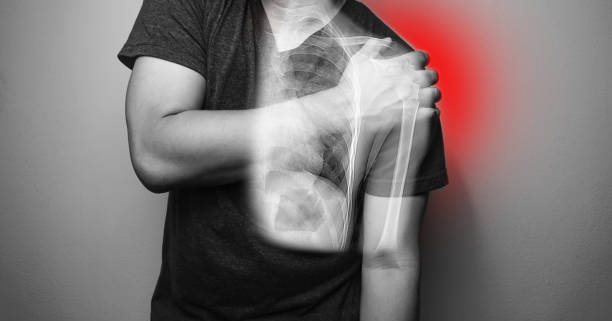
Shoulder dislocation can result from a range of activities, accidents, falls, or sports injuries. When your upper arm bone (humerus head) comes out of its socket (glenoid), it can damage the surrounding shoulder tissue, leading to significant pain and other issues. It’s crucial to reach out to a doctor promptly for proper treatment. In this blog, we will discuss the types, causes, symptoms, diagnosis, treatment, and ways to prevent shoulder dislocations. Let’s dive in!

Types of Shoulder Dislocation

Shoulder dislocations fall into three main categories, depending on the direction the upper arm bone (humerus) moves away from the socket joint after dislocation:

Anterior (Forward) Dislocation:

  • Most common type, making up over 95% of shoulder dislocation cases.
  • It typically results from an indirect force involving a combination of arm abduction, extension, and external rotation.
  • Anterior dislocation is frequently linked to injuries such as Labrum tears, significant tuberosity fractures, and humeral head fractures.

Posterior (Backwards) Dislocation:

  • Occurs when the humerus bone separates and moves away from the socket joint.
  • This type constitutes 2% to 4% of all shoulder dislocations.
  • Causes include seizures, electrical shocks, falling on an outstretched arm, or experiencing a blow to the front of the shoulder.

Inferior Dislocation:

  • The rarest type occurs in approximately 1 in every 200 cases.
  • Happens when the humerus is violently pushed downward.

Shoulder dislocations are usually a result of trauma, but in rare cases, they can occur with simple shoulder motions. These motions may be more likely to cause dislocations in individuals with weakened ligaments due to an inherent condition, potentially increasing the risk of dislocations in other joints.

It’s noteworthy that shoulder dislocations are the most common type of joint dislocations, primarily affecting young male adults and older women. This prevalence is because our upper extremities are extensively used in our daily activities, making shoulder joints more susceptible to dislocations compared to lower extremities.

Causes of Shoulder Dislocation

Shoulder dislocations can happen due to various reasons, especially during activities that involve intense force, twisting, or direct impact on the shoulder joint. Here are some common causes:

Forced Injury: A powerful blow to the shoulder or a sudden fall onto an outstretched arm can result in a shoulder dislocation. If the shoulder joint is twisted excessively, the ball of the upper arm bone may pop out of its socket.

Sports-related injury: Shoulder dislocation is a risk in contact sports such as football, rugby, and wrestling, as well as in sports like basketball and skiing.

Weak ligaments or tendon sprains: Individuals with weak or loose ligaments are more susceptible to shoulder dislocation because their joints lack the necessary stability.

Shoulder Dislocation Symptoms

Recognizing a shoulder dislocation involves observing several key signs:

Severe Pain: Movement intensifies the pain, and you may find it challenging to move your arm or experience complete immobility.

Inflammation and Bruising: Swelling and bruising around the shoulder joint are common indicators of a dislocation.

Deformed Appearance: The shoulder may look visibly out of place or deformed.

In addition to these, you might also experience numbness, tingling, or weakness near the injury. If you notice these shoulder dislocation symptoms, it’s crucial to seek immediate medical attention. While waiting for professional help, refrain from trying to relocate the shoulder yourself, as this could potentially harm nearby blood vessels, nerves, muscles, and ligaments. Using ice packs can help alleviate pain and reduce swelling.

Diagnosing Shoulder Dislocation

If you suspect any shoulder dislocation symptoms, it’s crucial to seek immediate medical attention. A healthcare professional will conduct a thorough assessment, which typically involves:

Physical Examination: The doctor will visually inspect the shoulder, assess its mobility, and inquire about your comfort level.

Imaging Tests: To identify injuries such as fractures or sprains, the medical expert may recommend X-rays, CT scans, or MRI tests.

Getting prompt medical help is essential for an accurate diagnosis and appropriate treatment.

Shoulder Dislocation Treatment

Addressing a shoulder dislocation involves various approaches:

Closed Reduction: A medical professional will gently maneuver the upper arm bone back into the shoulder socket. Sedation or anesthesia may be used for pain relief and muscle relaxation during this process.

Immobilization: After reduction, the shoulder may be immobilized for several weeks using a sling or brace. This helps promote healing and prevents further dislocations.

Surgery: For individuals with persistent instability or multiple dislocations, surgery may be recommended. Modern shoulder dislocation surgery is considered safe and effective, reducing the risk of re-injury, especially in young athletes.

Medication: Pain relievers and muscle relaxants will be prescribed to enhance comfort during the healing process.

Rehabilitation: After the immobilization period, comprehensive rehabilitation is essential. This involves exercises and physical therapy to restore strength, stability, and range of motion in the shoulder joint.

Preventing Shoulder Dislocations

Although some shoulder dislocations may be unavoidable, taking certain precautions can help minimize the risk:

Muscle Strengthening Exercises: Engage in exercises that strengthen shoulder muscles to improve stability and reduce the chances of dislocation.

Proper Equipment: When participating in activities or sports with intense physical contact, use appropriate gear such as shoulder pads or braces to provide added protection.

Correct Techniques: Whether exercising, playing sports, or lifting heavy weights, use proper techniques to minimize the risk of shoulder injury. It’s also essential to warm up before engaging in these activities.

Important Points to Remember

Shoulder dislocation is a painful condition that can significantly impact daily life and physical activity. Quick diagnosis and effective treatment are crucial for promoting healing and preventing repeated dislocations. To maintain the health of your shoulder joints, it’s important to be aware of the causes, shoulder dislocation symptoms, diagnosis, treatment, and preventive measures related to shoulder dislocation. If you suspect a shoulder dislocation, seek immediate medical care. Book your appointment today!

Clinical & Critical Cardiology

Clinical and Critical Cardiology: Understanding the Importance of Heart Health

Heart disease is the leading cause of death worldwide. According to the World Health Organization (WHO), an estimated 17.9 million people died from cardiovascular diseases (CVDs) in 2019, accounting for 32% of all global deaths. In the United States alone, heart disease is responsible for one in four deaths, making it the leading cause of death for both men and women.

Clinical and critical cardiology are two essential fields of medicine that deal with the prevention, diagnosis, and treatment of heart diseases. These two areas of cardiology are crucial in ensuring the optimal care of patients with heart conditions.

What is Clinical Cardiology?

Clinical cardiology is the branch of medicine that deals with the diagnosis and treatment of heart diseases. Clinical cardiologists specialize in the management of patients with various heart conditions, such as coronary artery disease, heart failure, valvular heart disease, and arrhythmias.

A clinical cardiologist typically evaluates patients who have symptoms of heart disease, such as chest pain, shortness of breath, palpitations, or fatigue. The cardiologist conducts a comprehensive evaluation, including a physical examination, medical history, and diagnostic tests, such as electrocardiogram (ECG), echocardiogram, stress test, or cardiac catheterization.

Based on the evaluation, the cardiologist provides a diagnosis and recommends an appropriate treatment plan, which may include medications, lifestyle modifications, or procedures such as angioplasty or heart surgery. The cardiologist also provides ongoing management and follow-up care to ensure that the patient’s heart condition is well-controlled and any potential complications are addressed promptly.

What is Critical Cardiology?

Critical cardiology is a specialized field of medicine that deals with the management of patients with acute or life-threatening heart conditions, such as acute myocardial infarction (heart attack), cardiogenic shock, or severe heart failure. Critical cardiologists work in intensive care units (ICUs) and provide round-the-clock care to patients who require close monitoring and advanced interventions.

Critical cardiologists are trained in advanced cardiac life support (ACLS) and are skilled in using various life-saving treatments and devices, such as mechanical ventilation, intra-aortic balloon pump, or extracorporeal membrane oxygenation (ECMO). These treatments help support the patient’s heart function and vital organs until the underlying condition is treated.

In addition to providing advanced interventions, critical cardiologists work closely with other healthcare professionals, such as cardiac surgeons, nurses, respiratory therapists, and pharmacists, to ensure the optimal care of critically ill patients. The goal is to stabilize the patient’s condition, minimize complications, and improve the chances of a full recovery.

Why is Heart Health Important?

Heart health is essential for overall health and well-being. The heart is a vital organ that pumps blood and oxygen to all parts of the body, providing the necessary nutrients and energy for cells to function properly. When the heart is not functioning correctly, it can lead to various complications, such as heart attack, stroke, kidney failure, or even death.

Maintaining a healthy heart involves adopting healthy lifestyle habits, such as regular exercise, a balanced diet, avoiding tobacco and excessive alcohol consumption, and managing stress. In addition, it is crucial to managing any underlying medical conditions that may affect heart health, such as hypertension, high cholesterol, or diabetes.

Early detection and treatment of heart disease are critical in preventing complications and improving outcomes. Regular check-ups with a cardiologist can help identify any potential risk factors or early signs of heart disease, allowing for prompt intervention and treatment.

Conclusion

Clinical and critical cardiology are two critical areas of medicine that play a crucial role in ensuring the optimal care of patients with heart diseases.

Paediatric Cardiology, Pediatrics, Cardiology, Heart Diseases, Congenital Heart Defects, Pediatric Cardiac Surgery, Pediatric Heart Conditions, Pediatric Cardiac Care.

Paediatric Cardiology: Understanding the Complexities of Children’s Heart Health

The heart is a crucial organ that pumps blood throughout the body, supplying vital oxygen and nutrients to all organs and tissues. Paediatric cardiology is a specialized field of medicine that focuses on the diagnosis, treatment, and management of heart disease in children from infancy to adolescence.

Children’s heart health is a complex subject that requires a deep understanding of both the anatomy and physiology of the heart, as well as the unique challenges that children face when it comes to heart health. In this blog post, we will explore some of the key aspects of paediatric cardiology, including common heart conditions in children, diagnostic procedures, and treatment options.

Common Heart Conditions in Children

Congenital Heart Defects

Congenital heart defects (CHD) are structural abnormalities that occur during the development of the heart and blood vessels in the womb. CHDs can range in severity from minor defects that may not require treatment, to life-threatening conditions that require immediate intervention.

Some of the most common types of CHDs include:

  • Atrial septal defects (ASD): A hole in the wall between the two upper chambers of the heart.
  • Ventricular septal defects (VSD): A hole in the wall between the two lower chambers of the heart.
  • Tetralogy of Fallot (ToF): A combination of four heart defects that affect blood flow through the heart and lungs.
  • Patent ductus arteriosus (PDA): Failure of a blood vessel that connects the aorta and pulmonary artery to close after birth.
  • Coarctation of the aorta: Narrowing of the aorta that restricts blood flow to the lower body.

Arrhythmias

Arrhythmias are irregular heart rhythms that can affect the heart’s ability to pump blood effectively. In children, arrhythmias can occur as a result of congenital heart defects, electrolyte imbalances, or underlying medical conditions.

Some common types of arrhythmias in children include:

  • Supraventricular tachycardia (SVT): A rapid heart rate that originates in the upper chambers of the heart.
  • Ventricular tachycardia (VT): A rapid heart rate that originates in the lower chambers of the heart.
  • Wolff-Parkinson-White syndrome (WPW): A rare condition where an extra electrical pathway in the heart can cause rapid heart rates.

Cardiomyopathy

Cardiomyopathy is a condition where the heart muscle becomes weak or stiff, making it difficult for the heart to pump blood effectively. In children, cardiomyopathy can be caused by genetic factors, viral infections, or other underlying medical conditions.

Some of the most common types of cardiomyopathy in children include:

  • Dilated cardiomyopathy: The heart becomes enlarged and weakened, reducing its ability to pump blood effectively.
  • Hypertrophic cardiomyopathy: The walls of the heart become thickened, making it harder for the heart to pump blood effectively.
  • Restrictive cardiomyopathy: The heart becomes stiff and unable to fill with blood properly.

Diagnostic Procedures in Paediatric Cardiology

Diagnosing heart conditions in children requires a thorough evaluation of the child’s medical history, physical examination, and a variety of diagnostic tests.

Some common diagnostic procedures in paediatric cardiology include:

  • Electrocardiogram (ECG): A test that records the electrical activity of the heart and can help identify abnormal heart rhythms or other electrical abnormalities.
  • Echocardiogram: A test that uses sound waves to create images of the heart and can help identify structural abnormalities or problems with the heart’s function.

Angioplasty is a minimally invasive procedure that is commonly performed in cardiology to treat coronary artery disease (CAD). The procedure involves using a balloon catheter to dilate narrowed or blocked coronary arteries, thereby restoring blood flow to the heart muscle. Angioplasty is often performed as part of a larger treatment plan for CAD, which may also include lifestyle changes, medication, or coronary artery bypass surgery. In this article, we will explore the different types of angioplasty, the indications for the procedure, and its associated risks and benefits.

Types of Angioplasty: There are several different types of angioplasty, each of which is used to treat different types of coronary artery disease. The most common types of angioplasty include:

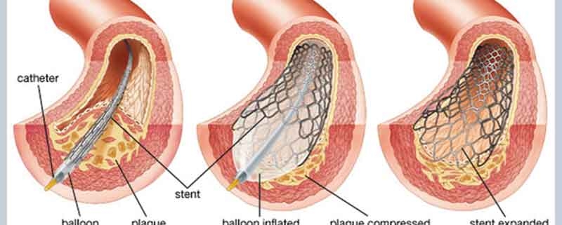
  1. Balloon angioplasty: Balloon angioplasty is the most common type of angioplasty. During this procedure, a small balloon is inserted into the blocked or narrowed coronary artery and inflated to widen the artery and restore blood flow.
  2. Drug-eluting stent: A drug-eluting stent is a small mesh tube that is inserted into the blocked or narrowed coronary artery. The stent is coated with a medication that is slowly released over time to prevent the artery from narrowing again.
  3. Coronary atherectomy: Coronary atherectomy is a procedure that involves using a small device to remove plaque from the walls of the coronary arteries. This procedure is often used in cases where the plaque is too hard or calcified to be treated with balloon angioplasty.

Indications for Angioplasty: Angioplasty is typically recommended for patients with significant blockages or narrowing of the coronary arteries. The procedure may be indicated in cases where the patient is experiencing symptoms such as chest pain, shortness of breath, or fatigue, which are often caused by reduced blood flow to the heart muscle.

Angioplasty may also be recommended for patients who have had a heart attack, as restoring blood flow to the affected area of the heart can help to prevent further damage and improve long-term outcomes. In some cases, angioplasty may also be recommended as a preventive measure for patients who are at high risk of developing CAD, such as those with a family history of the condition or those with multiple risk factors, such as high blood pressure, high cholesterol, or diabetes.

Risks and Benefits of Angioplasty: Like any medical procedure, angioplasty carries some risks, including bleeding, infection, and damage to the blood vessels or surrounding tissues. In rare cases, patients may experience more serious complications, such as heart attack, stroke, or kidney damage.

However, for most patients, the benefits of angioplasty far outweigh the risks. By restoring blood flow to the heart muscle, angioplasty can help to relieve symptoms, improve quality of life, and reduce the risk of heart attack and other complications. For many patients, angioplasty is an effective and relatively low-risk treatment option for coronary artery disease.

Conclusion: In conclusion, angioplasty is a common and effective treatment option for patients with coronary artery disease. The procedure is minimally invasive and can be performed on an outpatient basis, with most patients able to return to normal activities within a few days. While angioplasty carries some risks, for most patients, the benefits of the procedure far outweigh the potential complications. If you are experiencing symptoms of CAD or have been diagnosed with the condition, talk to your doctor about whether angioplasty may be a good treatment option for you.

Interventional Cardiology, Coronary Angioplasty, Stenting, Atherectomy, Laser Angioplasty, Rotational Atherectomy, Balloon Valvuloplasty, Transcatheter Aortic Valve Replacement, TAVR, Patent Foramen Ovale Closure, PFO Closure

Cardiovascular disease is one of the leading causes of death worldwide, and its management has been a major focus in modern medicine. Interventional cardiology is a subspecialty of cardiology that focuses on the treatment of heart disease using minimally invasive procedures. These procedures are designed to treat blockages in the coronary arteries that cause chest pain (angina) or a heart attack. In this blog, we will discuss interventional procedures in cardiology in detail.

Coronary Angioplasty:
Coronary angioplasty is a procedure that involves inserting a catheter (a long, thin tube) into the blocked artery and inflating a small balloon at the tip of the catheter to open up the blocked area. This is a minimally invasive procedure that is performed under local anaesthesia. The balloon is then deflated and the catheter is removed. In some cases, a stent (a small, mesh-like device) may be placed to keep the artery open. This procedure is commonly used to treat angina, heart attack, and other types of coronary artery disease.

Stenting:
Stenting is a procedure that involves inserting a stent into the blocked artery to keep it open. A stent is a small, mesh-like device that is placed inside the artery to prevent it from closing again. Stents can be made of different materials such as metal or plastic. This procedure is commonly used in conjunction with angioplasty.

Atherectomy:
Atherectomy is a procedure that involves removing plaque buildup from the walls of the arteries using a catheter. This procedure is usually performed in conjunction with angioplasty or stenting.

Laser Angioplasty:
Laser angioplasty is a procedure that uses a laser to remove plaque buildup from the walls of the arteries. This procedure is rarely used today due to the advent of other, more effective interventional procedures.

Rotational Atherectomy:
Rotational atherectomy is a procedure that involves removing plaque buildup from the walls of the arteries using a rotating catheter with a diamond tip. This procedure is usually performed in conjunction with angioplasty or stenting.

Balloon Valvuloplasty:
Balloon valvuloplasty is a procedure that involves inserting a catheter with a balloon at the tip into a narrowed heart valve. The balloon is then inflated to widen the valve opening. This procedure is commonly used to treat aortic stenosis, a condition in which the aortic valve narrows and restricts blood flow from the heart to the rest of the body.

Transcatheter Aortic Valve Replacement (TAVR):
Transcatheter aortic valve replacement (TAVR) is a minimally invasive procedure that involves replacing a diseased aortic valve with a new valve. The new valve is delivered to the heart through a catheter that is inserted into a blood vessel in the groin or chest. TAVR is typically performed in patients who are not good candidates for traditional open-heart surgery.

Patent Foramen Ovale (PFO) Closure:
Patent foramen ovale (PFO) closure is a procedure that involves closing a small hole in the heart that is present at birth. The hole, called the foramen ovale, is supposed to close shortly after birth but remains open in some people. This can cause blood clots to travel from the veins to the arteries, which can lead to a stroke. PFO closure involves inserting a catheter with a small device at the tip into the hole and deploying the device to close it.

In conclusion, interventional cardiology has revolutionized the treatment of heart disease by offering minimally invasive procedures that are safe and effective.