Cartilage Transplant Surgery in Dwarka, Delhi

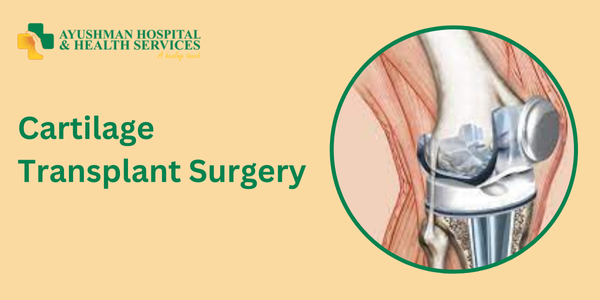
If you or someone you know is dealing with joint pain, limited movement, or a decrease in quality of life, cartilage transplant surgery might be a hopeful option. This innovative procedure aims to relieve pain and restore function in damaged joints. In this blog, we’ll walk you through everything you need to know about cartilage transplant surgery at Ayushman Hospital in Dwarka, Delhi. We’ll cover what cartilage is, why it gets damaged, the surgery details, recovery, and why many patients choose Ayushman Hospital for their care. Let’s dive in!

Introduction

Cartilage is a smooth, rubbery tissue that covers the ends of your bones in joints. Think of it as a cushion that helps your joints move smoothly and reduces friction. When cartilage gets damaged—due to injury, aging, or chronic conditions—it can lead to significant pain and limited mobility. Cartilage transplant surgery offers a way to replace damaged cartilage with healthy tissue, helping you regain movement and improve your quality of life.

At Ayushman Hospital in Dwarka, Delhi, patients benefit from advanced medical care and cutting-edge technology. With a team of skilled orthopedic surgeons and dedicated rehabilitation staff, the hospital is committed to helping you heal. This blog is designed to give you a clear understanding of the procedure and why it’s becoming a popular choice for many.

Understanding Cartilage and Its Importance

What is Cartilage?

Cartilage is a flexible yet tough tissue found in various parts of the body, including joints, the rib cage, ears, and nose. In your joints, it covers the ends of bones, allowing them to glide smoothly over one another and acting as a shock absorber. Unlike other tissues, cartilage doesn’t have its own blood supply, which makes it harder for it to heal itself when damaged.

The Role of Cartilage in Joint Function

  • Shock Absorption: Cartilage cushions the impact on your joints during activities like walking or running.
  • Smooth Movement: Its smooth surface allows bones to move with minimal friction.
  • Joint Stability: Healthy cartilage helps stabilize joints by evenly distributing weight.

When cartilage is damaged, it can cause pain, swelling, and reduced movement, leading to conditions like osteoarthritis that make daily activities difficult.

Causes of Cartilage Damage

Understanding what leads to cartilage damage can help you take preventive measures. Here are some common causes:

Traumatic Injuries

Accidents, falls, or sports injuries can cause sudden damage to cartilage. For example, twisting your knee during a game can lead to a cartilage tear, resulting in immediate pain and swelling.

Degenerative Conditions

Over time, wear and tear on your joints can degrade cartilage. Osteoarthritis is a common condition where cartilage wears away, causing chronic pain and limited mobility.

Genetic Factors

Some people are genetically predisposed to cartilage damage. While you can’t change your genes, early diagnosis and treatment can help manage symptoms effectively.

Inflammatory Diseases

Conditions like rheumatoid arthritis can cause joint inflammation, leading to cartilage breakdown over time.

Lifestyle Factors

  • Obesity: Extra weight adds stress to your joints, speeding up cartilage wear.
  • Sedentary Lifestyle: Not exercising regularly can weaken muscles and joints, affecting cartilage health.
  • Poor Nutrition: A diet lacking essential nutrients can impair your body’s ability to maintain healthy cartilage.

To prevent further damage, it’s important to adopt a healthy lifestyle, eat well, and seek medical advice if you experience joint pain.

What is Cartilage Transplant Surgery?

Cartilage transplant surgery is a procedure designed to repair or replace damaged cartilage. The goal is to restore joint function, reduce pain, and improve your overall quality of life.

Types of Cartilage Transplant Procedures

Several types of procedures exist, and the right one for you depends on the severity of your cartilage damage:

  • Autologous Chondrocyte Implantation (ACI): Healthy cartilage cells are taken from you, grown in a lab, and then re-implanted into the damaged area.
  • Osteochondral Autograft Transfer (OAT): A small plug of healthy cartilage and bone is taken from a non-weight-bearing part of your joint and placed in the damaged area.
  • Osteochondral Allograft Transplantation: For larger areas of damage, donor tissue is used to replace the defective cartilage.

How Does the Surgery Work?

The process typically involves these steps:

  1. Assessment and Planning: Imaging studies (like an MRI) are done to assess the damage, and the surgeon plans the best approach.
  2. Harvesting the Cartilage: Depending on the procedure, your own cartilage or donor cartilage is prepared.
  3. Implantation: The damaged cartilage is removed, and the healthy cartilage is transplanted into the area.
  4. Stabilization: Sometimes, devices are used to stabilize the new tissue during healing.

The surgery is usually performed under general anesthesia, and the time it takes can vary based on the complexity of the case.

Who Can Benefit from the Surgery?

Cartilage transplant surgery can be a transformative option, but it’s not for everyone. Here’s who might benefit:

Ideal Candidates

  • Young to Middle-Aged Adults: Generally, those who are active and have localized cartilage defects benefit the most.
  • Isolated Cartilage Damage: Those with damage confined to one area of the joint, without affecting other structures, are excellent candidates.
  • Good Overall Health: Individuals who maintain a healthy lifestyle and do not have underlying health issues are preferred.

Who May Not Benefit?

  • Advanced Joint Disease: Patients with widespread arthritis may not see significant improvement.
  • Older Adults with Multiple Health Issues: Recovery may be more challenging for older patients with various health concerns.
  • Unrealistic Expectations: It’s important to understand that while the surgery can improve function and relieve pain, it may not fully restore the joint.

A thorough evaluation by an orthopedic specialist at Ayushman Hospital can help determine if this surgery is right for you.

Preoperative Evaluation and Preparation

Before your surgery, you’ll undergo a thorough evaluation at Ayushman Hospital to ensure the best outcomes.

Medical History and Physical Examination

The process starts with a review of your medical history and a physical exam to assess joint function and overall health.

Diagnostic Imaging

Advanced imaging techniques (like MRI or X-rays) are used to:

  • Determine the extent of cartilage damage.
  • Identify underlying issues like bone spurs.
  • Plan the precise location and size of the cartilage defect.

Consultation with Specialists

At Ayushman Hospital, you’ll consult with a team of specialists, including orthopedic surgeons, radiologists, and rehabilitation experts, to create a comprehensive care plan.

Preoperative Instructions

You’ll receive detailed instructions to prepare for surgery, including:

  • Medications: Guidelines on which medications to stop or continue.
  • Fasting: Information on fasting before the operation.
  • Lifestyle Modifications: Recommendations for diet and activity to promote optimal healing.

This thorough evaluation helps ensure you’re ready for a safe and effective surgery.

The Surgical Procedure in Detail

Let’s take a closer look at what happens during the surgery:

Step 1: Anesthesia

You’ll be given anesthesia to keep you comfortable and pain-free during the procedure.

Step 2: Making the Incision

Once the anesthesia takes effect, the surgeon makes a small incision near the affected joint.

Step 3: Removal of Damaged Cartilage

The surgeon carefully removes the damaged cartilage, creating a clean area for the new cartilage.

Step 4: Harvesting Healthy Cartilage

  • Autologous Procedure: A small sample of your own cartilage is taken from a non-critical area and processed in a lab.
  • Allograft Procedure: If donor tissue is used, healthy cartilage is sourced from a donor.
Step 5: Implanting the New Cartilage

The surgeon carefully implants the healthy cartilage into the prepared site, ensuring it fits well with the surrounding area.

Step 6: Stabilization and Closure

Once the new cartilage is in place, the surgeon may use small devices or sutures to secure it. The incision is then closed.

Step 7: Final Assessment

Before concluding the surgery, the team checks that the implant is stable and that everything is properly aligned.

The entire process can take a few hours, depending on the complexity of your case.

Post-Surgery Recovery and Rehabilitation

After your surgery, recovery is a gradual process that involves several stages. Here’s what you can expect:

Immediate Postoperative Care

  • Monitoring in Recovery: You’ll be closely monitored as you wake up from anesthesia. The medical staff will check your vital signs and manage any immediate discomfort.
  • Pain Management: Pain medications will be given to keep you comfortable.

Hospital Stay

Your hospital stay can vary:

  • Short Stay: Some patients can go home within 24 to 48 hours.
  • Extended Stay: More complex cases may require a longer stay for observation.

Rehabilitation and Physical Therapy

Rehabilitation is essential for a successful recovery. Your physical therapy program will help:

  • Restore Joint Function: Exercises will improve mobility, flexibility, and strength.
  • Promote Healing: Gentle movements encourage blood flow to the joint.
  • Prevent Complications: Structured rehabilitation helps avoid issues like stiffness and muscle weakness.

Long-Term Recovery

  • Gradual Increase in Activity: You’ll gradually start increasing your activity levels, following your therapist’s guidelines.
  • Regular Follow-Ups: Keeping up with your orthopedic surgeon for follow-up visits is important for tracking your progress.
  • Lifestyle Adjustments: Eating well and staying active will support your joint health long-term.

Successful recovery can take several months, but with commitment, many patients experience significant improvements in their joint function and a reduction in pain.

Success Rates and Expected Outcomes

Many patients see positive results from cartilage transplant surgery, leading to improved mobility and a better quality of life. However, individual outcomes can vary based on several factors:

Factors Affecting Success

  • Extent of Damage: The size and location of the cartilage defect matter.
  • Age and Health: Younger, healthier patients usually heal better.
  • Adherence to Rehabilitation: Following your rehab program closely is critical.
  • Surgical Expertise: The experience of the surgical team plays a big role in success.

What Patients Can Expect

  • Pain Reduction: Most patients report less pain after recovery.
  • Improved Mobility: Better joint function allows you to return to everyday activities.
  • Enhanced Quality of Life: With reduced pain and improved movement, many feel a boost in their overall well-being.

Long-Term Outlook

While cartilage transplant surgery may not completely eliminate joint issues, it can significantly delay the need for more invasive procedures like joint replacement. Regular care and a healthy lifestyle are key to maintaining the benefits of your surgery.

Why Choose Ayushman Hospital, Dwarka, Delhi?

Ayushman Hospital is known for its expertise in advanced surgical procedures, including cartilage transplant surgery. Here are some reasons why patients choose this hospital:

Experienced Surgical Team

The hospital has a team of skilled orthopedic surgeons who specialize in joint surgeries. Their experience ensures you receive top-notch care.

State-of-the-Art Facilities

Ayushman Hospital is equipped with modern diagnostic and surgical tools, allowing for precise imaging and advanced surgical techniques.

Personalized Care

Every patient gets individualized attention. The team works closely with you to understand your unique needs, from evaluation to rehabilitation.

Comprehensive Postoperative Rehabilitation

The hospital has a dedicated rehabilitation unit where physical therapists design tailored recovery plans to help you regain full functionality.

Patient-Centric Approach

From your first consultation to follow-up visits, Ayushman Hospital prioritizes your comfort and understanding throughout the entire process.

Strategic Location

Located in Dwarka, Delhi, the hospital is easily accessible, making it a convenient option for patients in the area.

Patient Experiences and Testimonials

Hearing from others who have undergone this surgery can be inspiring. Many patients at Ayushman Hospital have shared their success stories:

  • A New Lease on Life: One young athlete described how the surgery helped him return to sports after years of knee pain.
  • Effective Pain Management: Many patients report a significant reduction in pain, allowing them to resume daily activities with ease.
  • Improved Joint Function: Testimonials often highlight increased mobility and stability, enabling a more active lifestyle.

These experiences show that with the right care, cartilage transplant surgery can lead to a remarkable improvement in quality of life.

Frequently Asked Questions (FAQs)

  1. What is the main purpose of cartilage transplant surgery?
    • The surgery aims to repair or replace damaged cartilage, reducing pain and improving mobility.
  2. How long is the recovery process?
    • Recovery can vary, but expect several weeks for initial healing and up to six months for full rehabilitation.
  3. Is the surgery painful?
    • While some pain is expected, modern pain management techniques help minimize discomfort.
  4. Who is an ideal candidate for this surgery?
    • Ideal candidates are usually younger to middle-aged adults with localized cartilage damage and good overall health.
  5. How do I prepare for the surgery?
    • Preparation includes a thorough medical evaluation, imaging, and consultations with the surgical team.
  6. Are there any risks involved?
    • Potential risks include infection and incomplete cartilage integration, but careful planning helps minimize these.
  7. Can the surgery completely restore joint function?
    • While the surgery can significantly reduce pain and improve function, it may not fully restore the joint.

The Journey to a Healthier Joint

Cartilage transplant surgery is more than just a procedure—it represents a journey toward a better, more active life. Deciding to undergo surgery is a big step, and it’s important to weigh the benefits and risks carefully.

A Step Towards Recovery

For many, the surgery is the first step in reclaiming independence. It offers the chance to return to activities that were once limited by pain. The journey begins with understanding cartilage damage and progresses through well-planned medical and rehabilitation steps.

A Collaborative Effort

Successful outcomes rely on teamwork between the patient, surgical team, and rehabilitation specialists. At Ayushman Hospital, this collaborative approach ensures you receive continuous care from the moment you decide to have surgery until you’ve fully recovered.

Embracing a New Lifestyle

After surgery, adopting a lifestyle that supports joint health is key. Regular exercise, a balanced diet, and routine check-ups can help maintain your joint health and overall well-being.

Looking Forward

The field of cartilage transplant surgery is advancing rapidly. At Ayushman Hospital, ongoing improvements in surgical techniques and postoperative care are helping more patients enjoy better outcomes. There’s a bright future ahead for those seeking relief from joint pain.

Conclusion

Cartilage transplant surgery at Ayushman Hospital in Dwarka, Delhi, offers a modern solution for those struggling with joint pain. With advanced surgical techniques, personalized care, and comprehensive rehabilitation, the hospital is dedicated to restoring joint function and enhancing quality of life.

This guide has covered:

  • The importance of cartilage for joint health.
  • Common causes of cartilage damage.
  • Different types of cartilage transplant procedures.
  • The surgical process and recovery journey.
  • Factors that influence success and expected outcomes.
  • Why Ayushman Hospital is a trusted choice for this procedure.

If you’re considering cartilage transplant surgery, consulting with an orthopedic specialist is crucial. The path to better joint health is a collaborative one, and with the expertise at Ayushman Hospital, you can take confident steps toward a more active, pain-free life.

If you have further questions or want to schedule a consultation, don’t hesitate to reach out to Ayushman Hospital. Their dedicated team is ready to assist you every step of the way.

Thank you for reading this guide on cartilage transplant surgery. We hope it has provided valuable insights and answered your questions. Remember, your journey to better joint health is just a consultation away!

Book an Appointment Today!

🌐 Visit: www.ayushmanhhs.in

Would you like more information on costs, insurance, or doctor recommendations?

Bankart Repair Surgeries

Understanding the Shoulder and the Bankart Lesion

The Anatomy of Your Shoulder

Your shoulder is one of the most flexible joints in the body. It is designed to move in multiple directions, allowing you to reach, throw, and lift. This mobility is made possible by a ball-and-socket structure, where the rounded head of the upper arm bone (humerus) fits into a shallow socket in the shoulder blade (glenoid). A rim of cartilage called the labrum surrounds the socket and acts as a cushion, stabilizing the joint during movement.

What is a Bankart Lesion?

A Bankart lesion is a specific injury to the labrum, usually occurring when the shoulder dislocates. When the shoulder pops out of place, the labrum may tear from the edge of the glenoid, leading to persistent instability. This injury is most common in young athletes or individuals who have experienced repeated shoulder dislocations due to sports injuries, accidents, or trauma. If left untreated, a Bankart lesion can lead to chronic pain, repeated dislocations, and difficulty with daily tasks.

What is Bankart Repair Surgery?

The Goal of the Surgery

Bankart repair surgery aims to reattach the torn labrum to the glenoid and tighten any stretched ligaments. This repair helps restore the stability of the shoulder, reducing the chance of future dislocations. There are two primary surgical approaches:

  • Arthroscopic Repair: This minimally invasive procedure uses small incisions, a camera, and specialized instruments to repair the labrum. Because of the smaller incisions, patients usually experience less pain and a quicker recovery.
  • Open Repair: In some cases, particularly if the injury is severe, an open surgery might be required. This traditional approach involves a larger incision to access the shoulder joint directly. While recovery might be slightly longer, open repair can be very effective for complex cases.

How Does the Surgery Help?

Bankart repair surgery not only fixes the torn labrum but also helps restore the natural anatomy of the shoulder. This repair is essential for patients who have experienced recurrent shoulder dislocations because it reinforces the joint and minimizes the risk of future injuries. By stabilizing the shoulder, the surgery helps reduce pain and improves overall shoulder function, allowing patients to return to their daily activities and sports with confidence.

When is Bankart Repair Surgery Needed?

Recognizing the Signs

Not everyone with a shoulder injury needs surgery. However, if you have suffered repeated shoulder dislocations or experience ongoing pain and instability, your doctor might recommend a Bankart repair. Common signs that indicate surgery might be necessary include:

  • Recurrent Dislocations: If your shoulder frequently pops out of place even after treatment or physical therapy.
  • Persistent Pain: Continuous discomfort that does not improve with non-surgical treatments.
  • Limited Range of Motion: Difficulty performing everyday tasks or sports activities due to shoulder weakness or stiffness.
  • Physical Instability: A feeling that the shoulder is “loose” or unstable during movement.

Who is a Good Candidate?

Candidates for Bankart repair surgery often include:

  • Athletes: Especially those involved in contact sports or activities that require overhead movements.
  • Active Individuals: Anyone who values a high level of physical activity and needs a stable shoulder for their lifestyle.
  • Patients with Recurrent Injuries: Individuals who have experienced multiple dislocations and have not found relief through conservative treatments.

Before recommending surgery, doctors at Ayushman Hospital conduct thorough evaluations, including physical examinations and imaging studies like MRI or CT scans, to determine the exact nature and severity of the injury.

Bankart Repair Surgery at Ayushman Hospital, Dwarka, Delhi

About Ayushman Hospital

Located in the heart of Dwarka, Delhi, Ayushman Hospital is renowned for its commitment to patient care and advanced medical treatments. The hospital has built a reputation for excellence in various specialties, including orthopedics and sports medicine. With state-of-the-art facilities, a team of highly skilled surgeons, and a patient-centered approach, Ayushman Hospital is well-equipped to handle complex procedures like Bankart repair surgery.

The Expert Team

At Ayushman Hospital, the orthopedic department is led by experienced surgeons who have performed numerous Bankart repair procedures. Their expertise in both arthroscopic and open techniques means that each patient receives personalized care tailored to their specific condition. The team works closely with physiotherapists, pain management specialists, and nursing staff to ensure a smooth surgical process and a speedy recovery.

Advanced Facilities and Technology

Ayushman Hospital boasts modern operating theaters and advanced diagnostic tools. The use of cutting-edge imaging technology allows surgeons to assess the injury with precision. During the surgery, high-definition cameras and specialized instruments assist in minimally invasive procedures, reducing recovery time and minimizing discomfort for patients.

A Patient-Centered Approach

Understanding that surgery can be a daunting experience, Ayushman Hospital places great emphasis on patient comfort and support. From the initial consultation through the recovery process, patients are guided by a compassionate team dedicated to answering questions, addressing concerns, and ensuring that every step of the journey is as smooth as possible.

The Surgical Procedure in Detail

Pre-Surgical Evaluation

Before undergoing Bankart repair surgery, patients undergo a comprehensive evaluation. This evaluation includes:

  • Medical History and Physical Examination: The surgeon reviews your health history and conducts a physical exam to assess shoulder stability and pain.
  • Imaging Tests: X-rays, MRI, or CT scans are used to get a clear picture of the shoulder anatomy and to confirm the presence and extent of a Bankart lesion.
  • Discussion of Options: The surgeon will discuss whether an arthroscopic or open repair is most suitable based on the severity of the injury and your lifestyle needs.

The Day of Surgery

On the day of the surgery, patients are admitted to Ayushman Hospital’s surgical unit. Here is an outline of what to expect:

  1. Anesthesia: Patients are given either general anesthesia or regional anesthesia with sedation, ensuring a pain-free procedure.
  2. Positioning: Once asleep, you will be positioned comfortably on the operating table with special supports to stabilize the shoulder.
  3. Incisions: For an arthroscopic procedure, small incisions (often less than 1 cm) are made around the shoulder. These incisions allow the insertion of a small camera and surgical instruments.
  4. Visualization: The camera provides a clear view of the inside of your shoulder, enabling the surgeon to locate the torn labrum and assess the damage accurately.
  5. Repair Process: Using tiny instruments, the surgeon carefully reattaches the labrum to the glenoid bone. Anchors and sutures may be used to secure the tissue in place.
  6. Closure: Once the repair is complete, the incisions are closed with small stitches. For open surgery, the process is similar but involves a larger incision and more direct access to the joint.
  7. Recovery in Hospital: Patients are monitored in the recovery room until the anesthesia wears off, and vital signs are stable.

Duration of the Procedure

The entire surgery typically takes between one to two hours, depending on the complexity of the tear and the chosen surgical method. In most cases, patients can go home the same day or after a short hospital stay.

Benefits of Bankart Repair Surgery

Restoring Shoulder Stability

One of the primary benefits of Bankart repair surgery is the restoration of shoulder stability. By reattaching the torn labrum and tightening the surrounding ligaments, the surgery reduces the likelihood of future dislocations, giving patients a more secure and stable joint.

Reducing Pain and Discomfort

Chronic shoulder instability often leads to persistent pain. By addressing the root cause of the instability, Bankart repair surgery can significantly reduce or eliminate this pain, making daily activities more comfortable.

Improving Quality of Life

A stable shoulder allows patients to return to their favorite activities, whether it’s sports, exercise, or simply playing with their children. The improved function can lead to a better quality of life, increased confidence, and a renewed sense of freedom.

Shorter Recovery Times

Thanks to advancements in minimally invasive techniques, many patients experience shorter recovery times with arthroscopic Bankart repair compared to traditional open surgery. This means less time away from work, sports, and social activities.

Preventing Further Damage

Repeated shoulder dislocations can cause additional wear and tear on the joint, potentially leading to arthritis or other complications. By repairing the damage early, Bankart repair surgery helps prevent further deterioration of the shoulder joint.

Recovery and Rehabilitation Process

Immediate Post-Surgery Care

After the surgery, patients are moved to a recovery area where they are closely monitored. It is normal to experience some pain and swelling, which can be managed with prescribed medications and ice packs. The hospital staff at Ayushman Hospital ensures that you are comfortable and well-informed about your post-operative care.

Physical Therapy and Rehabilitation

A key component of a successful recovery is physical therapy. The rehabilitation process generally includes:

  • Initial Immobilization: For the first few weeks, the shoulder may be placed in a sling or brace to keep it immobile and allow the tissues to heal.
  • Gentle Exercises: Once healing has begun, a physiotherapist will introduce gentle exercises to restore range of motion without straining the repaired tissue.
  • Strengthening Exercises: Gradually, more intensive exercises are added to strengthen the muscles around the shoulder, providing better support for the joint.
  • Gradual Return to Activity: Over several months, patients are guided on how to safely return to daily activities and sports.

Recovery Timeline

The recovery timeline can vary depending on the severity of the injury and the type of surgery performed. Here is a general guide:

  • 0–2 Weeks: Focus on pain management and reducing swelling. The shoulder is typically immobilized.
  • 2–6 Weeks: Begin gentle range-of-motion exercises under the guidance of a physiotherapist.
  • 6–12 Weeks: Gradually introduce strengthening exercises.
  • 3–6 Months: Most patients notice significant improvement in shoulder stability and function. Full return to sports and high-impact activities may be possible after this period.

Ongoing Support

Ayushman Hospital provides ongoing support throughout the recovery process. Follow-up appointments ensure that the healing process is on track and allow the medical team to address any concerns promptly. Patients also receive detailed instructions on exercises and lifestyle modifications to protect their shoulder in the long term.

Patient Experiences and Success Stories

Many patients who have undergone Bankart repair surgery at Ayushman Hospital share inspiring stories of recovery and renewed activity. Consider the story of Ravi, a young athlete who struggled with recurrent shoulder dislocations. Ravi’s condition forced him to sit out of sports and limited his everyday activities. After a detailed consultation and evaluation at Ayushman Hospital, he underwent an arthroscopic Bankart repair. With a dedicated rehabilitation program and the continuous support of his medical team, Ravi was soon back to playing sports, enjoying his hobbies, and leading a more active life. His journey is a testament to the transformative impact of modern surgical techniques and personalized patient care.

The Role of Advanced Technology and Expert Surgeons

Cutting-Edge Surgical Tools

Ayushman Hospital is proud to use the latest surgical technology, which plays a crucial role in the success of Bankart repair surgeries. High-definition cameras, precision instruments, and advanced imaging techniques allow the surgeons to perform the procedure with great accuracy. This precision not only reduces the risk of complications but also minimizes trauma to the surrounding tissues, leading to faster recovery times.

A Team of Dedicated Experts

The success of any surgery depends largely on the expertise of the surgical team. At Ayushman Hospital, each patient benefits from the knowledge and experience of surgeons who have performed hundreds of orthopedic procedures. Their familiarity with both arthroscopic and open techniques ensures that the chosen method is tailored to the patient’s specific needs. This level of expertise is supported by a dedicated team of anesthesiologists, nursing staff, and physiotherapists who work together to provide comprehensive care.

Emphasis on Patient Safety

Patient safety is at the forefront of every procedure at Ayushman Hospital. The hospital follows strict protocols to ensure that every surgery is performed in a sterile environment with the highest standards of care. Regular training sessions, state-of-the-art operating theaters, and continuous monitoring contribute to a safe surgical experience for every patient.

Frequently Asked Questions About Bankart Repair Surgery

What is a Bankart lesion, and why does it occur?

A Bankart lesion is a tear of the labrum in the shoulder, typically caused by a dislocation. It often occurs in individuals who experience repeated shoulder dislocations, especially athletes and people with high levels of physical activity.

How is Bankart repair surgery performed?

Bankart repair can be performed using minimally invasive arthroscopic techniques or via open surgery. The procedure involves reattaching the torn labrum to the glenoid bone and tightening the ligaments to restore shoulder stability.

How long is the recovery period?

Recovery varies from patient to patient, but generally, the initial healing phase takes about 2–6 weeks, with full rehabilitation and return to sports or high-impact activities occurring between 3–6 months.

Will I experience pain after the surgery?

Some pain and discomfort are normal after surgery. However, the medical team at Ayushman Hospital provides effective pain management strategies to ensure a smooth recovery.

Are there any risks associated with Bankart repair surgery?

As with any surgical procedure, there are risks such as infection, nerve injury, or complications related to anesthesia. However, these risks are minimized by the hospital’s experienced team and advanced surgical techniques.

How do I know if I’m a good candidate for Bankart repair surgery?

Your orthopedic surgeon will evaluate your shoulder’s condition through physical examinations and imaging tests. If you have recurrent dislocations and persistent instability that does not improve with non-surgical treatments, you may be a good candidate for the surgery.

What should I expect on the day of the surgery?

On the day of surgery, you will undergo anesthesia, and the surgical team will carefully prepare you for the procedure. The surgery itself typically lasts one to two hours, followed by a period of recovery in the hospital’s postoperative unit.

Conclusion

Bankart repair surgery is a highly effective treatment for patients suffering from recurrent shoulder dislocations and instability. By repairing the torn labrum and restoring the natural anatomy of the shoulder, the procedure offers significant relief from pain, improves shoulder stability, and enhances overall quality of life. Whether you are an athlete eager to return to sports or someone looking to regain confidence in your daily movements, Bankart repair can provide the lasting solution you need.

Ayushman Hospital in Dwarka, Delhi, stands out as a beacon of excellence in orthopedic care. With advanced facilities, cutting-edge technology, and a team of experienced surgeons, the hospital is dedicated to ensuring that every patient receives personalized care and achieves the best possible outcome. From the initial consultation and detailed pre-surgical evaluation to the expertly performed procedure and comprehensive rehabilitation program, every step of your journey is managed with care and professionalism.

If you or a loved one has been struggling with shoulder instability, it may be time to consider Bankart repair surgery. The dedicated team at Ayushman Hospital is ready to guide you through the process, answer your questions, and provide the support you need for a successful recovery. With the right treatment and care, you can look forward to a future filled with improved mobility, reduced pain, and the freedom to enjoy the activities you love.

Book an Appointment Today!

🌐 Visit: www.ayushmanhhs.in

Would you like more information on costs, insurance, or doctor recommendations?


Bladder Cancer

Bladder cancer, while not the most common form of cancer, often goes unnoticed due to a lack of public awareness. This can lead to delayed diagnosis and hinder treatment success. This blog aims to empower you with knowledge about bladder cancer, including its signs and symptoms, the importance of early detection, and various treatment options available at Ayushman Hospital & Health Services.

Understanding Bladder Cancer

Bladder cancer begins in the cells that line the bladder, the organ that holds urine. Transitional cell carcinoma (TCC), also known as urothelial carcinoma, is the most prevalent type. TCC can be non-invasive, confined to the inner bladder layer, or invasive, spreading deeper into the bladder wall. Other types that are less frequently seen include squamous cell carcinoma and adenocarcinoma.

Warning Signs and Symptoms

Bladder cancer often presents with several telltale signs, although it’s important to remember that these can also indicate other conditions. Here’s what to watch out for:

  • Blood in the Urine (Hematuria): This is a common symptom, with urine appearing pink, red, or cola-colored. While hematuria can have various causes, including infections or kidney stones, prompt investigation is crucial, especially if it recurs.
  • Urinary Changes: You may experience increased urination frequency, urgency to urinate, or difficulty passing urine despite feeling the need. These can mimic a urinary tract infection (UTI) but persist despite treatment.
  • Painful Urination: Burning or discomfort during urination can be a sign of bladder cancer, UTIs, or other bladder conditions. Seek medical evaluation if it persists.
  • Pelvic or Lower Back Pain: As the cancer progresses, discomfort or pain may develop in the pelvic region or lower back, particularly if the cancer spreads to nearby tissues or organs.
  • Weight Loss and Fatigue: Advanced bladder cancer can lead to unintentional weight loss and fatigue, often indicating a more developed stage where the cancer may have spread.
  • Bone Pain: If the tumor spreads (metastasizes) to bones, pain can occur, particularly in the pelvis, spine, or long bones.

Early Detection Saves Lives

Early detection is paramount for successful bladder cancer treatment for several reasons:

  • More Treatment Options: Early-stage cancer is usually confined to the inner bladder lining, allowing for less invasive procedures like cystoscopy with tumor removal or transurethral resection of bladder tumor (TURBT). These offer high success rates and positive long-term outcomes.
  • Improved Prognosis: Early diagnosis significantly increases the chances of a complete cure. When detected and treated early, the likelihood of successful treatment and long-term survival is much higher. Conversely, delayed diagnosis allows the cancer to progress, making treatment more challenging and reducing the probability of a favorable outcome.
  • Preserving Bladder Function: Early intervention can often preserve bladder function. In advanced stages, removing the entire bladder (cystectomy) might be necessary, significantly impacting quality of life.
  • Enhanced Quality of Life: Early detection not only improves the chances of successful treatment but also allows individuals to maintain a better quality of life throughout their treatment journey. By addressing the tumor in its early stages, individuals may experience fewer symptoms and have a shorter recovery time, enabling them to resume normal activities quicker.

Treatment Options

Ayushman Hospital & Health Services offers a comprehensive range of bladder cancer treatment options tailored to individual needs, considering factors like cancer stage and grade, overall health, and patient preferences. Here’s an overview of the available treatments:

  • Surgery:
    • Transurethral Resection of Bladder Tumor (TURBT): This minimally invasive procedure is the most common for early-stage bladder cancer. A thin, lighted instrument is inserted through the urethra to reach the bladder, where the surgeon uses an electrical current to remove the tumor. TURBT can also be used for diagnosis.
    • Radical Cystectomy: This surgical procedure entails removing the entire bladder, along with adjacent lymph nodes and nearby organs. It’s typically recommended for invasive bladder cancer that has spread beyond the inner lining. After surgery, a urinary diversion is created, such as an ileal conduit (using a part of the small intestine) or a neobladder (a pouch from a nearby intestine) to allow urine to leave the body.
  • Chemotherapy: Chemotherapy medications can be used before or after surgery (neoadjuvant or adjuvant chemotherapy) for muscle-invasive bladder cancer (MIBC) to shrink tumors, reduce recurrence risk, or treat advanced cancer. In some cases, it may be the primary treatment for individuals who cannot undergo surgery.
  • Immunotherapy: Immunotherapy drugs are increasingly used for advanced or metastatic bladder cancer that has not responded well to other treatments. These medications work by stimulating the body’s immune system to target and destroy cancer cells.
  • Radiation Therapy: Radiation therapy employs high-energy X-rays or other types of radiation to eliminate cancer cells or reduce tumor size. There are two main types used for bladder cancer:
  • External Beam Radiation Therapy: This type is the most frequently encountered. A large machine outside the body directs beams of radiation precisely at the tumor and surrounding tissues.
  • Internal Beam Radiation Therapy (Brachytherapy): In some cases, a radioactive implant may be placed directly into the bladder for a short period. This approach delivers a concentrated dose of radiation to the tumor while minimizing exposure to healthy tissues.

Conclusion

Bladder cancer is a treatable condition, especially when detected early. By understanding the signs and symptoms, and taking a proactive approach to your health, you can significantly improve your chances of a successful outcome. If you experience any concerning symptoms, don’t hesitate to seek medical advice at Ayushman Hospital & Health Services. Timely diagnosis and treatment are essential for achieving the best results. Remember, understanding empowers you to manage your health proactively. Share this information with your loved ones and encourage them to prioritize regular checkups and screenings.

Impact of Acute Respiratory Failure on Lung Health

Acute respiratory failure (ARF) is a life-threatening condition where the lungs struggle to deliver enough oxygen to the bloodstream and remove excess carbon dioxide. This vital gas exchange is compromised, leading to organ damage and the urgent need for medical attention. ARF can develop suddenly (acute) or worsen gradually from pre-existing breathing problems (chronic). Understanding the causes, risk factors, and consequences of ARF is crucial for protecting lung health and seeking timely intervention.

Causes of Acute Respiratory Failure: A Multifaceted Threat

Several medical conditions can trigger ARF, each posing a unique threat to lung function:

  • Pneumonia: Severe lung infections cause inflammation and fluid buildup in the air sacs, hindering oxygen absorption.
  • Chronic Obstructive Pulmonary Disease (COPD): Long-term respiratory illnesses like COPD can worsen, leading to acute respiratory failure.
  • Pulmonary Embolism: Blockages in the pulmonary arteries restrict blood flow to the lungs, hindering oxygen exchange.
  • Acute Respiratory Distress Syndrome (ARDS): Severe lung injury due to trauma or infections causes extensive inflammation, impacting breathing.
  • Asthma: Severe asthma attacks can drastically restrict airways, limiting gas exchange and airflow.
  • Neuromuscular Disorders: Diseases like muscular dystrophy and ALS weaken respiratory muscles, making breathing difficult.
  • Inhalation Injuries: Inhaling smoke, fumes, vomit, or experiencing near-drowning can cause acute lung injury and respiratory failure.
  • Sepsis: Widespread inflammation from this life-threatening infection can extend to the lungs, causing respiratory failure.
  • Head/Chest Injuries: Severe trauma to the head, chest, or other areas can directly damage the lungs or the brain’s respiratory center, impacting breathing.

Risk Factors: Who’s More Susceptible to ARF?

Certain factors increase the likelihood of developing ARF, particularly in individuals with existing health issues:

  • Chronic Lung Conditions: Those with COPD, asthma, or other chronic lung ailments are at higher risk.
  • Age: Elderly individuals are more susceptible due to weakened respiratory function.
  • Premature Birth: Premature babies with underdeveloped lungs, pulmonary hypertension, or lung birth defects are at higher risk.
  • Smoking: Damages lung tissue and weakens the immune system, increasing ARF risk.
  • Obesity: Excess weight can restrict lung expansion and hinder breathing.
  • Weakened Immune System: Compromised immunity due to drugs, chronic illnesses, or HIV/AIDS increases ARF risk.
  • Sedatives: Certain medications used during surgery can suppress breathing, leading to ARF, especially in high-risk individuals.
  • Occupational Hazards: Exposure to chemicals, dust, and pollutants at work can damage the lungs and raise ARF risk.
  • Physical Inactivity: Lack of exercise weakens heart and lung function, increasing ARF risk.

The Devastating Consequences of ARF on Lung Health

ARF’s impact on lung health is severe, with both immediate and long-term effects:

  • Tissue Damage: Low blood oxygen and high carbon dioxide levels cause tissue hypoxia (oxygen deprivation) and inflammation. This can damage lung tissue and lead to fibrosis (scarring).
  • Strained Respiratory Muscles: Difficulty breathing forces patients to exert more energy to stay ventilated, straining respiratory muscles and potentially worsening their condition.
  • Increased Infection Risk: Mechanical ventilation can increase the risk of secondary infections, adding to patients’ health challenges.
  • Higher Mortality Risk: Patients with a history of ARF, especially those with recurrent episodes or underlying chronic conditions, have a higher risk of death.

Treatment Strategies: Fighting for Recovery

Due to its potentially fatal nature, ARF requires immediate medical diagnosis and emergency care in a hospital setting. Treatment focuses on stabilizing the patient, addressing the underlying cause, and supporting the lungs until they can function independently. Key components of ARF treatment include:

  • Oxygen Therapy: Supplemental oxygen is administered to maintain adequate blood oxygen levels.
  • Mechanical Ventilation: When patients cannot breathe adequately on their own, mechanical ventilation may be necessary. This can involve non-invasive techniques like masks or invasive techniques like endotracheal intubation.
  • Medications: Depending on the cause, treatment may involve diuretics to reduce fluid buildup, bronchodilators to open airways, antibiotics to fight infections, and corticosteroids to reduce inflammation.
  • Treating the Underlying Cause: Addressing the root cause of ARF, such as treating infections or managing flare-ups of chronic diseases, is crucial for recovery.
  • Supportive Care: Proper nutrition, hydration, and monitoring are essential, alongside physical therapy and pulmonary rehabilitation to strengthen respiratory muscles and improve lung function.

Preventing ARF: Taking Charge of Your Lung Health

The key to preventing ARF lies in managing underlying conditions and reducing risk factors. Here are some essential strategies:

  • Regular Medical Checkups and Treatment Adherence: Schedule regular checkups with your doctor to monitor your health and identify any potential respiratory issues early on. If you have a chronic lung condition like COPD or asthma, diligently follow your doctor’s treatment plan, which may include medications, inhalers, and lifestyle modifications. This proactive approach can help prevent flare-ups and minimize the risk of ARF.
  • Vaccination: Getting vaccinated against respiratory infections like influenza and pneumonia is crucial, especially for those at higher risk, such as the elderly and individuals with chronic health conditions. These vaccinations significantly reduce the chances of developing severe respiratory illnesses that could progress to ARF.
  • Smoking Cessation: Smoking is a major risk factor for ARF. Quitting smoking is one of the most effective ways to protect your lungs and reduce your risk of developing respiratory problems, including ARF. Numerous resources and support programs are available to help you quit smoking successfully.
  • Maintaining a Healthy Weight: Obesity can strain your lungs and make breathing difficult. Maintaining a healthy weight through a balanced diet and regular exercise can significantly improve lung function and reduce the risk of ARF.
  • Healthy Lifestyle Habits: A healthy lifestyle that incorporates regular physical activity, a balanced diet rich in fruits, vegetables, and whole grains, and adequate sleep is essential for overall health, including lung function. Exercise strengthens your respiratory muscles and improves lung capacity, making you less susceptible to respiratory problems.
  • Occupational Safety: If your work environment involves exposure to dust, chemicals, or fumes, wear appropriate personal protective equipment (PPE) like masks and respirators to minimize lung damage. Talk to your employer about workplace safety measures to protect your respiratory health.
  • Environmental Awareness: Be mindful of air quality, especially if you live in an area with high pollution levels. Limit outdoor activity on days with high pollution and consider using air purifiers indoors to improve air quality.

By implementing these preventive measures and maintaining a healthy lifestyle, you can significantly reduce your risk of developing ARF and safeguard your lung health. Remember, early detection and intervention are critical for successful ARF treatment. If you experience any symptoms of respiratory distress, such as shortness of breath, rapid breathing, chest tightness, or wheezing, seek immediate medical attention.

Hyperthyroidism in Men

Men and thyroid problems? It might sound uncommon, but an overactive thyroid, or hyperthyroidism, can significantly impact men’s health. This condition often whispers before it roars, with subtle signs that can be easily overlooked. This blog dives deep into hyperthyroidism in men, exploring its causes, symptoms, diagnosis, treatment, and when to seek medical help.

Why Does My Thyroid Act Up?

The thyroid gland, located in your neck, is like a body’s master controller, regulating metabolism, energy, and hormones. When it goes rogue, producing too much thyroid hormone, hyperthyroidism disrupts everything.

Here’s what can trigger this imbalance in men:

  • Autoimmune Attack: Graves’ disease, the most common culprit, tricks your immune system into attacking your thyroid, revving it up.
  • Family Ties: A family history of thyroid issues raises your risk.
  • Too Much or Too Little Iodine: Iodine is essential for thyroid function, but too much or too little can cause problems.
  • Hidden Culprits: Certain medical conditions and even growths on the thyroid can affect hormone production.

Symptoms: Don’t Ignore the Whispers

Hyperthyroidism’s symptoms can be sneaky, mimicking other health concerns or stress. Here’s what to watch out for:

  • Shrinking Waistline: Unexpected weight loss despite normal eating.
  • Heat Wave: Feeling constantly hot and sweaty.
  • Racing Heart: Rapid heartbeat and palpitations.
  • Neck Lump: A swollen thyroid gland (goiter).
  • Brain Fog: Difficulty concentrating or remembering things.
  • Mood Swings: Feeling anxious, irritable, or on edge.
  • Muscle Matters: Weakness and fatigue.
  • Bedroom Blues: Reduced sex drive or erectile dysfunction.
  • Bathroom Blues: Changes in bowel habits (diarrhea or constipation).
  • Hair Loss: Thinning hair or hair loss.
  • Dry Skin and Brittle Nails
  • Sleepless Nights: Trouble falling asleep or staying asleep.
  • Rarely: Breast enlargement (gynecomastia)

Getting a Diagnosis

Early diagnosis is key to managing hyperthyroidism. Doctors use a combination of approaches:

  • Checking Your Neck: A physical exam to assess thyroid size and signs of increased metabolism.
  • Blood Tests: Measuring hormone levels (TSH, T3, T4) to identify imbalances.
  • Antibody Tests: Looking for specific antibodies in autoimmune cases (like Graves’ disease).
  • Imaging Tests: Scans or ultrasounds to visualize the thyroid gland for abnormalities.
  • Biopsy (if needed): A tissue sample examination to rule out cancer in case of nodules.

Treatment Options: Taking Back Control

The treatment plan depends on the severity, cause, and your overall health. Here are the main approaches:

  • Medications: Antithyroid drugs can help regulate hormone production. Careful monitoring is crucial to avoid overcorrection or undercorrection.
  • Radioactive Iodine (RAI) Therapy: A targeted approach using radioactive iodine to destroy overactive thyroid cells. This may lead to hypothyroidism (underactive thyroid), requiring lifelong hormone replacement therapy.
  • Surgery: In some cases, removing part or all of the thyroid gland might be necessary. This is a definitive solution but typically results in hypothyroidism, requiring hormone replacement therapy.
  • Managing Symptoms: Medications or lifestyle changes can address specific symptoms like rapid heart rate, weight issues, fatigue, or sexual dysfunction.

Final Notes

Watch out for warning signs like unexplained weight changes, persistent mood swings, or irregular heartbeats. Early diagnosis and proper treatment are crucial for managing hyperthyroidism effectively. If you notice any concerning symptoms or have a family history of thyroid problems, visit the doctor.

Ayushman Hospital & Health Services is a state-of-the-art facility with a team of highly trained experts dedicated to providing exceptional care for various health conditions, including thyroid problems. Schedule your appointment today!

Different Types of Bone Fractures

Bone fractures, often a result of falls or accidents, encompass a wide spectrum of injuries. They can be as subtle as hairline cracks or as severe as shattered bones. To ensure proper healing and optimal recovery, understanding the different fracture types and their corresponding treatment options is essential. This article delves into the complexities of bone fractures, exploring various fracture classifications, common types, causes, diagnosis, treatment approaches, and the importance of follow-up care.

Understanding Bone Fractures

A bone fracture simply refers to a broken or cracked bone due to various reasons, like falls, accidents, or underlying medical conditions that weaken bones.

Classifying Bone Fractures

There are several ways to categorize bone fractures based on different characteristics. Here are four key classifications:

  • Open vs. Closed Fractures: Open fractures, also called compound fractures, involve the broken bone protruding through the skin or creating an open wound, increasing the risk of infection. Closed fractures, or simple fractures, don’t pierce the skin, reducing the infection risk.
  • Displaced vs. Non-Displaced Fractures: Displaced fractures occur when the bone fragments are misaligned, requiring manipulation or surgery for realignment and stabilization. In non-displaced fractures, the bone is broken but the fragments remain relatively intact, potentially leading to a quicker healing process.

Common Types of Bone Fractures

  • Transverse Fracture: A straight break occurring perpendicular to the bone’s long axis, often caused by a direct blow.
  • Spiral Fracture: Caused by a twisting force, the fracture line spirals around the bone shaft, resembling a staircase. Common in sports injuries and car accidents.
  • Greenstick Fracture: Unique to children with flexible bones, a greenstick fracture involves a bend and partial break, resembling a green stick that bends but doesn’t snap completely.
  • Stress Fracture: Tiny cracks or hairline fractures caused by repetitive stress or overuse, common in athletes and individuals engaged in high-impact activities.
  • Compression Fracture: The collapse of a vertebra, often due to osteoporosis or trauma, causing pain, height loss, and curvature of the spine.
  • Other Fracture Types: Additional fractures include oblique fractures (angled break), impacted fractures (one fragment driven into another), segmental fractures (multiple breaks), and comminuted fractures (bone shatters into multiple fragments).

Symptoms of a Bone Fracture

Symptoms can vary depending on the location and severity of the fracture, but common signs include:

  • Pain, especially at the injury site, worsens with movement or pressure.
  • Swelling and bruising around the injured area due to inflammation and bleeding.
  • Visible deformity or misalignment in severe fractures.
  • Difficulty or inability to move the affected limb or joint.
  • Tenderness to the touch in the area around the fracture.
  • Numbness, tingling, or weakness below the injury site if nerves are affected.
  • Bone popping sound (crepitus), when broken bone ends, rub together.
  • Visible bone protrusion in open fractures.

What Causes Bone Fractures?

  • Trauma: Falls, sports injuries, car accidents, or direct blows to the bone.
  • Repetitive Stress: Overuse or repetitive stress on a bone can lead to stress fractures.
  • Osteoporosis: Weakens bones, making them more susceptible to fractures, especially in the spine, hips, and wrists.
  • Pathological Conditions: Certain medical conditions like bone cancer, bone diseases, or infections can weaken bone structure and increase fracture risk.
  • Age: Older adults are more prone due to decreased bone density, muscle mass, and impaired balance and coordination.
  • Sports and Activities: High-impact activities like football or gymnastics increase the risk due to physical demands and potential falls or collisions.
  • Accidents: High-speed collisions can cause severe injuries, including multiple fractures.
  • Violence: Fractures can also result from intentional acts of violence.

Diagnosing Bone Fractures

Diagnosis typically involves a detailed medical history, physical examination, and imaging studies:

  • Medical History: The doctor asks about the injury, symptoms, and any pre-existing medical conditions.
  • Physical Examination: The doctor examines the affected area for tenderness, swelling, deformity, and changes in range of motion.
  • Imaging Studies: X-rays are most common, followed by CT scans for complex fractures, MRIs for soft tissue injuries, bone scans for suspected but not visible fractures, and ultrasounds in specific cases.

Treatment Options for Bone Fractures

Treatment depends on the fracture type, location, displacement, patient’s age and health, and activity level. Here are some common approaches:

  • Immobilization: The primary goal is to stabilize the fractured bone for proper healing using casts, splints, braces, or traction. Immobilization helps prevent further injury, reduces pain, and allows the bone to heal correctly in the proper alignment.
  • Reduction: If the fracture is displaced, it may need realignment, a procedure called reduction. This can be done in two ways:
    • Closed Reduction: Performed without surgery, a doctor manipulates the bone fragments back into their correct position.
    • Open Reduction: Surgery is required to achieve proper alignment and stability. This may involve internal fixation using screws, plates, or rods, or external fixation using pins or screws placed outside the skin to hold the bones in place.
  • Medications: Pain relievers like over-the-counter or prescription medications may be recommended to manage pain during healing. Additionally, medications to prevent infection or promote bone healing may be prescribed.
  • Physical Therapy: Once healing begins, physical therapy exercises are crucial to restore strength, flexibility, and range of motion in the affected area. These exercises help prevent muscle atrophy, improve function, and facilitate a return to normal activities.
  • Follow-up Care: Regular appointments with your healthcare provider are essential to monitor healing progress, assess treatment effectiveness, and make adjustments if needed. X-rays or other imaging studies may be repeated to ensure proper bone alignment.

Conclusion

Bone fractures can vary significantly in severity. However, with proper diagnosis, treatment, and preventive measures, most fractures heal completely, and individuals can regain full function. Early intervention is crucial for optimal healing and preventing future complications. If you have concerns about your bone health or have experienced a fracture, consult a healthcare professional for proper diagnosis and guidance.

Laparoscopic Surgery

Traditionally, surgery has been associated with anxiety, pain, and lengthy recovery times. However, advancements in medical technology have introduced minimally invasive procedures like laparoscopic surgery, transforming the surgical landscape.

What is Laparoscopic Surgery?

Laparoscopic surgery, also known as minimally invasive surgery (MIS) or keyhole surgery, utilizes specialized instruments and a camera called a laparoscope. Tiny incisions are made, and the laparoscope transmits magnified images from inside the body, allowing surgeons to operate with exceptional precision. At Ayushman Hospital & Health Services, we with our team of highly skilled surgeons and cutting-edge equipment, offer effective laparoscopic procedures for a wide range of conditions, enhancing patients’ quality of life.

Applications of Laparoscopic Surgery

Laparoscopic surgery’s versatility extends to various medical specialties, including:

  • General Surgery: This minimally invasive approach is suitable for hernia repair, gallbladder removal, appendectomy, splenectomy, and treatment of various colon conditions.
  • Gynecology: Laparoscopy aids in diagnosing and treating endometriosis, performing tubal sterilization for permanent birth control, removing benign ovarian cysts, and treating uterine conditions, including cancer.
  • Urology: The procedure can address prostate cancer, bladder issues, and even remove a diseased kidney.
  • Gastroenterology: Laparoscopic surgery can be used for liver tumor or cyst removal, treating severe GERD (Gastroesophageal Reflux Disease), and performing weight-loss procedures.
  • Other Specialties: Laparoscopic techniques can enhance outcomes in some thoracic surgeries, joint surgeries, and spine procedures. It’s also used to treat conditions like pyloric stenosis, choledochal cysts, and undescended testes in children.

Advantages of Laparoscopic Surgery

While smaller incisions and faster recovery times are widely recognized benefits, laparoscopic surgery offers a multitude of lesser-known advantages that make it a preferred choice for patients:

  1. Enhanced Cosmetic Outcome: Ayushman Hospital’s skilled laparoscopic surgeons leave behind minimal, barely noticeable scars. This significantly improves cosmetic outcomes, boosting patients’ confidence as they return to their daily lives.
  2. Reduced Hernia Risk: Smaller incisions and minimal tissue trauma during laparoscopy significantly reduce the risk of postoperative hernias compared to traditional open surgeries.
  3. Improved Lung Function: Innovative laparoscopic techniques used in thoracic surgeries lead to a noticeable improvement in lung function after surgery, especially beneficial for patients with pre-existing lung conditions.
  4. Better Tissue Preservation: Ayushman Hospital’s laparoscopic surgeons utilize precise surgical movements, allowing for the preservation of healthy tissues surrounding the surgical site. This is particularly crucial in cancer surgeries for improved long-term outcomes.
  5. Less Postoperative Fatigue: Compared to traditional surgeries, laparoscopic procedures minimize postoperative fatigue and exhaustion, enabling patients to resume daily activities and work much quicker.
  6. Lower Risk of Adhesions: Laparoscopic surgeries significantly reduce the formation of adhesions (scar tissue) within the abdomen. This decreases the risk of bowel obstructions and future complications.
  7. Lower Risk of Infection: The procedure minimizes the exposure of internal organs to external contaminants, leading to a significantly reduced risk of postoperative infections.

Final Note

While laparoscopic surgery has some potential side effects, its minimally invasive nature, smaller incisions, reduced pain, faster recovery, and shorter hospital stay make it the preferred choice for treating various conditions. Ayushman Hospital is committed to providing exceptional care through our team of expert surgeons. We offer effective laparoscopic surgeries at affordable prices, aiming to improve your quality of life.

Gastric Cancer: Causes, Symptoms, Diagnosis & Treatment

Gastric cancer, also known as stomach cancer, is a serious health condition that arises when abnormal cells in the stomach lining multiply uncontrollably, forming a tumor. These malignant cells can spread to other parts of the body through the bloodstream or lymphatic system if left untreated. This article delves into the complexities of gastric cancer, exploring its various types, risk factors, diagnosis methods, and available treatment approaches.

Understanding Gastric Cancer

The inner lining of the stomach is responsible for producing digestive juices that break down food. Gastric cancer disrupts this process by causing the uncontrolled growth of abnormal cells within this lining. The vast majority of stomach cancers (around 90-95%) are adenocarcinomas, originating from the glandular cells that secrete digestive enzymes.

Who Faces a Higher Risk?

Certain factors can increase an individual’s susceptibility to gastric cancer. These include:

  • Age: The risk of developing gastric cancer rises significantly with age, particularly for those above 60 years old.
  • Family History: Having a close relative with a history of stomach cancer elevates your risk.
  • Stomach Conditions: Pre-existing stomach issues like ulcers can contribute to an increased risk.
  • Lifestyle Habits: Smoking tobacco and consuming excessive amounts of alcohol are significant risk factors.
  • Dietary Choices: Diets high in processed meats, smoked foods, and salted foods, coupled with a lack of fruits and vegetables, can contribute to the development of gastric cancer.
  • Helicobacter Pylori (H. pylori) Infection: This bacterial infection can cause chronic inflammation in the stomach lining, potentially leading to cancer in some cases.

It’s important to remember that even individuals without these risk factors can develop gastric cancer. Early detection is crucial for a favorable prognosis.

The Different Faces of Gastric Cancer

Gastric cancer manifests in various forms, each with its own characteristics:

  • Adenocarcinomas: As mentioned earlier, these are the most common type of stomach cancer. They can be further classified into two subtypes:
    • Intestinal Type: This subtype generally has a better prognosis and may respond well to targeted therapy due to specific genetic traits. It tends to grow slower and stay localized within the stomach for a longer duration before spreading.
    • Diffuse Type: This type spreads more aggressively compared to the intestinal type. It’s less common but presents treatment challenges due to its rapid growth and metastasis.
  • Gastrointestinal Stromal Tumours (GISTs): These are a less frequent type of sarcoma arising from specific cells located throughout the digestive system. Their behavior can vary, with some exhibiting slow growth and others spreading rapidly to the liver and surrounding tissues.
  • Neuroendocrine Tumours: These tumors develop from cells with features of both nerve and hormone-producing cells. While some are slow-growing, others can progress quickly. Diagnosis and treatment depend on the tumor’s grade and stage.
  • Lymphomas: These cancers originate from lymphocytes, a type of immune cell. Lymphomas of the stomach, such as diffuse large B-cell lymphoma and MALT lymphoma, require specific treatment approaches like chemotherapy and immunotherapy.

Recognizing the Warning Signs

Early-stage gastric cancer often doesn’t present with noticeable symptoms. However, as the cancer progresses, some potential signs and symptoms may emerge, including:

  • Indigestion-like pain: This discomfort can occur in the upper middle abdomen, similar to ulcer pain, due to stomach acid irritating the inflamed cancerous tissue.
  • Swallowing difficulties: A sensation of food getting stuck in the throat can indicate a tumor obstructing the passage of food into the stomach.
  • Persistent heartburn or acid reflux: A growing tumor can hinder the movement of food through the digestive tract, leading to frequent heartburn.
  • Digestive issues: Bloating, nausea, loss of appetite, and feeling full after small meals are common as tumor growth disrupts normal digestion and stomach emptying.
  • Unexplained weight loss: This can occur as the body burns extra calories due to cancer inflammation or struggles to absorb nutrients due to the spread of cancer.
  • Fatigue and weakness: These symptoms often develop as the body combats the disease and experiences reduced energy intake due to appetite loss.
  • Vomiting blood: Blood in vomit, appearing black in color, suggests internal bleeding from the cancerous lesion in the upper gastrointestinal tract.
  • Dizziness: Low iron levels caused by chronic blood loss can lead to dizziness.
  • Abdominal swelling: Fluid buildup around the stomach due to metastases releasing fluids into surrounding tissues can cause swelling.
  • Skin problems: Advanced cases, where cancer cells spread through the bloodstream, can manifest as unusual skin bumps or changes.

Causes of Gastric Cancer and Exploring Treatment Options

While the exact causes of gastric cancer remain under investigation, researchers have identified several contributing factors:

  • Dietary Habits: Regularly consuming processed meats, smoked foods, and salted foods can damage the stomach lining over time. Additionally, a diet lacking in antioxidants, found in fruits and vegetables, can also contribute to the risk.
  • H. pylori Infection: Chronic infection with this bacterium can cause chronic inflammation in the stomach, potentially leading to cell mutations and cancer development.
  • Genetic Predisposition: Having a close family member with gastric cancer increases your risk. Specific gene mutations can also be hereditary factors.
  • Anatomical Changes: Prior surgeries like partial stomach removal and conditions like pernicious anemia can elevate the risk.
  • Lifestyle Choices: Smoking tobacco, excessive alcohol consumption, obesity, and physical inactivity are modifiable risk factors.
  • Occupational Exposures: Prolonged contact with certain substances like coal dust, rubber dust, asbestos, and specific metals may contribute to the risk.
  • Radiation Exposure: Prior radiation therapy for other cancers can increase the risk of developing gastric cancer.
  • Oncogene Imbalances: Genetic alterations that accelerate cell growth, such as KRAS mutations and HER2 gene amplification, can play a role in tumor progression.

By minimizing controllable risk factors through healthy lifestyle choices and regular screenings, individuals can potentially reduce their chances of developing gastric cancer.

Diagnosing Gastric Cancer: A Multifaceted Approach

Early and accurate diagnosis is crucial for effective treatment of gastric cancer. Physicians typically employ a combination of diagnostic tests, including:

  • Endoscopy: A thin, flexible tube with a camera (endoscope) is inserted through the mouth to examine the digestive tract. Tissue samples can be collected during the procedure for further analysis.
  • Endoscopic Ultrasound: This specialized technique combines endoscopy with ultrasound technology to provide detailed images of the stomach wall layers, aiding in the detection of deeper tumors.
  • Barium Swallow: The patient drinks a liquid containing barium while X-ray images are captured to visualize the throat and stomach for abnormalities.
  • CT Scan: This imaging technique uses X-rays and computer technology to generate detailed cross-sectional images of the stomach, aiding in tumor detection and staging. Contrast agents may be administered orally and intravenously to enhance image clarity.
  • PET Scan: This test involves injecting a mildly radioactive sugar solution into the bloodstream. Cancer cells are known to take up more sugar than healthy cells. A PET scan can detect these areas of increased sugar uptake, potentially revealing the presence and spread of cancer.
  • Laparoscopy: A thin, lighted tube is inserted through a small incision in the abdomen to visually examine the abdominal cavity and nearby organs for signs of metastasis.
  • Blood Tests: These tests can help identify anemia and other potential indicators of gastric cancer.
  • Stool Test: This test checks for the presence of blood in the stool, which could be a sign of upper gastrointestinal bleeding.
  • Biopsy: This definitive diagnostic procedure involves extracting a small tissue sample from the suspicious area. The sample is then examined under a microscope to confirm the presence or absence of cancer cells and determine the cancer type.

Tailoring Treatment to the Individual: A Look at Treatment Options

The chosen treatment approach for gastric cancer depends on various factors such as the type and stage of the cancer, the patient’s overall health, and available treatment options:

  • Surgery: This is the primary treatment for most curable gastric cancers. It may involve removing all or part of the stomach (gastrectomy) along with nearby lymph nodes. Laparoscopic techniques can sometimes be employed, offering quicker recovery times.
  • Supportive (Palliative) Care: This approach focuses on improving the patient’s quality of life and managing symptoms such as pain, nausea, and fatigue, particularly in advanced or incurable cases.
  • Chemotherapy: This treatment utilizes powerful drugs delivered intravenously to destroy cancer cells. It is often used before surgery to shrink the tumor or after surgery to reduce the risk of recurrence.
  • Targeted Therapy: For advanced cancers with specific genetic mutations, targeted drugs can be used to attack these specific molecular pathways, potentially offering a more precise approach to treatment.
  • Immunotherapy: This relatively new approach involves stimulating the body’s immune system to recognize and fight cancer cells. Immunotherapy has shown promising results in treating gastric cancer, offering hope for future advancements.
  • Radiation Therapy: Focused high-energy X-rays are used to kill or shrink tumors. It can be used in combination with other therapies or for palliative purposes to manage pain in advanced stages.

Conclusion

Gastric cancer, though a serious condition, can be effectively managed with early detection and proper treatment. By understanding the different types, risk factors, symptoms, and available treatment options, individuals can empower themselves to empower themselves to make informed decisions about their health. Early detection through regular checkups and awareness of potential symptoms is crucial for a favorable prognosis. Additionally, adopting a healthy lifestyle that incorporates a balanced diet, regular exercise, and maintaining a healthy weight can significantly reduce the risk of developing gastric cancer. While ongoing research continues to unveil more about the complexities of this disease, advancements in diagnosis and treatment offer a brighter outlook for those battling gastric cancer. Remember, knowledge is power, and arming yourself with information is the first step towards taking control of your health.

How to Keep Your Spine Safe from a Disc Prolapse

The human spine is a marvel of engineering, providing both support and flexibility. However, this complex structure can be susceptible to injury, including disc prolapse, also known as a slipped disc. This condition occurs when the soft inner core of a spinal disc bulges or ruptures through the tougher outer ring, potentially pressing on nearby nerves and causing pain, numbness, or weakness.

Understanding the anatomy of the spine and the causes of disc prolapse is crucial for taking preventative measures.

What is Disc Prolapse?

Disc prolapse happens when the outer ring of a disc weakens or tears, allowing the inner core to protrude. This protrusion can compress neighboring nerves, leading to a variety of symptoms. The location of the prolapsed disc determines the specific nerves affected and the resulting symptoms.

Symptoms of Disc Prolapse

A disc prolapse may manifest in any spinal region, though it typically emerges in the lower back (lumbar spine) or occasionally in the neck (cervical spine). Symptoms fluctuate based on the disc’s location and the nerves affected. The following are among the most frequently encountered symptoms:

  • Pain: This is the most frequent symptom, often described as sharp, burning, or electric shock-like. Pain can radiate from the lower back down the buttocks and leg (sciatica) if the disc prolapse is in the lumbar spine. In the cervical spine, pain may travel into the shoulder and arm. Certain movements, coughing, sneezing, or maintaining specific positions can worsen the pain.
  • Numbness and Tingling: People with a slipped disc often experience a pins-and-needles sensation or numbness in the area served by the affected nerve.
  • Weakness: Muscles controlled by the compressed nerves may become weak, leading to difficulty walking, stumbling, or problems holding or lifting objects.
  • Loss of Bladder or Bowel Control: In severe cases, the nerves controlling bladder and bowel function can be affected, leading to loss of control. This is a rare symptom.

Causes of Disc Prolapse

Disc prolapse isn’t caused by the disc slipping out of place. Instead, it’s the bulging or herniation of the inner material that creates problems. Several factors can contribute to disc prolapse:

  • Disc Degeneration: As we age, the discs in our spine naturally lose water content and flexibility, making them more susceptible to tears or ruptures. This is the most common cause of disc prolapse.
  • Injury: A sudden impact, such as a fall or forceful twisting of the spine, can cause a disc to herniate.
  • Physical Activities: Certain activities that put excessive strain on the spine, like repetitive bending, heavy lifting, or forceful twisting, can increase the risk of disc prolapse. Jobs requiring constant pulling, pushing, or lifting can also be risk factors.
  • Genetics: Some people have a genetic predisposition to weaker disc material, making them more susceptible to prolapse.
  • Lifestyle Choices: Smoking, obesity, and excessive stress can accelerate disc degeneration and increase the risk of prolapse.

Treatment for Disc Prolapse

The good news is that most disc prolapse cases resolve on their own within 6-8 weeks. Treatment usually focuses on pain management and promoting healing. Here are some common treatment options:

  • Medication: Non-prescription pain relievers such as ibuprofen or acetaminophen are effective in alleviating mild to moderate pain. Corticosteroid injections near the affected nerves may be used for more severe pain. Muscle relaxants may be prescribed to address muscle spasms.
  • Physiotherapy: Physiotherapy exercises can strengthen core muscles, improve flexibility, and reduce pain. A physiotherapist will teach you specific exercises and postures to manage your disc prolapse.
  • Home Remedies: Applying hot or cold packs to the affected area can provide temporary pain relief and reduce inflammation. Staying active with light walks or gentle stretching is recommended to prevent muscle stiffness. However, avoid prolonged bed rest. Remember to maintain proper form and avoid sudden movements while bending or lifting.
  • Surgery: In severe cases where conservative treatment fails to provide relief for several weeks, surgery may be recommended. Surgery typically involves removing only the protruding portion of the disc. In rare cases, the entire disc may be removed, and the vertebrae may be fused for stability.

Preventing Disc Prolapse: Taking Charge of Your Spinal Health

Disc prolapse can be a debilitating condition, but the good news is that there are several steps you can take to significantly reduce your risk. Here are some key strategies for keeping your spine healthy and preventing disc prolapse:

  • Embrace Regular Exercise: Physical activity strengthens the muscles that support your spine, improving its overall stability and reducing pressure on the discs. Aim for regular exercise that incorporates both strength training and flexibility exercises. Engaging in swimming, walking, yoga, and Pilates can be fantastic options.
  • Maintain Good Posture: Poor posture, especially slouching, puts excessive strain on the discs in your lower back. Remember to maintain good posture all day long, whether you’re seated, standing, or on the move. Here are some tips:
    • Keep your shoulders back and relaxed.
    • Keep the natural arch in your lower back.
    • When sitting, ensure your feet are flat on the floor and knees bent at a 90-degree angle.
    • Consider investing in a supportive ergonomic chair with excellent lumbar support.
  • Lift Smart: Lifting heavy objects is a common cause of disc prolapse. Here’s how to lift safely:
    • Bend your knees and maintain a straight back while lifting.
    • Engage your core muscles for support.
    • If the object feels too heavy, don’t hesitate to request assistance.
  • Maintain a Healthy Weight: Excess weight puts extra stress on your spine and discs. Losing weight or maintaining a healthy weight can significantly reduce your risk of disc prolapse.
  • Quit Smoking: Smoking weakens the discs and hinders the healing process. Quitting smoking is not only beneficial for your overall health but also for your spinal health.
  • Manage Stress: Chronic stress can contribute to muscle tension and tightness, which can put strain on your spine. Techniques like meditation, yoga, and deep breathing can help manage stress and promote relaxation.
  • Listen to Your Body: Be attentive to the signals it sends. If you experience any pain or discomfort in your back, especially after strenuous activity, take a break and rest. Could you please provide more context or clarify what you’d like rewritten? I’m here to help!
  • Consider Chiropractic Care: While not a cure-all, chiropractic adjustments can improve spinal alignment and joint function, potentially reducing the risk of disc prolapse. However, it’s important to consult with your doctor before starting chiropractic care.

Conclusion

By incorporating these preventative measures into your lifestyle, you can significantly reduce your risk of developing disc prolapse. Remember, a healthy spine is essential for maintaining mobility, preventing pain, and ensuring an active life. If you experience any persistent back pain or suspect a disc prolapse, consult a healthcare professional for proper diagnosis and treatment. Early intervention can prevent the condition from worsening and ensure a faster recovery.

Brain Tumor

Experiencing frequent headaches can be concerning, especially if they worsen over time. While headaches are a common occurrence, they can sometimes be an indicator of an underlying condition. This article explores frequent headaches, brain tumors, and the warning signs to look out for.

Understanding Frequent Headaches

Headaches classified as chronic or frequent occur for 15 or more days each month over three months. These headaches can be mild or severe and significantly disrupt daily life. Some common types of chronic headaches include:

  • Tension headaches: These headaches feel like a tight band around the head.
  • Migraines: These are intense, throbbing headaches that can affect one or both sides of the head and may be accompanied by aura, a tingling sensation, or visual disturbance.
  • Cluster headaches: These headaches come and go in cycles, causing severe pain on one side of the head, usually around or behind the eye.
  • Hemicrania continua: This is a constant, daily headache on one side of the head, often resembling a migraine.
  • New daily persistent headache: This type of headache involves a sudden onset of daily pain that persists for months without relief.

Brain Tumors and How They Cause Headaches

Brain tumors are abnormal growths of cells within the brain. They can be benign (non-cancerous) or malignant (cancerous). Brain tumors can disrupt normal brain function and cause various health problems, including headaches.

Headaches caused by brain tumors differ from regular headaches. These headaches arise due to the tumor’s pressure on surrounding tissues and the disruption of normal brain function. The specific characteristics of the headache can vary depending on the tumor’s location and size. Here are some common signs:

  • Pain nature: The pain can be dull and persistent or sharp and throbbing, depending on the tumor’s location.
  • Morning headaches: These headaches are often worse upon waking due to fluid buildup in the brain during sleep.
  • Activity-induced worsening: Activities that increase pressure in the head, such as bending over, coughing, or lifting heavy objects, can worsen the headache.
  • Resistance to medication: Unlike regular headaches that respond to over-the-counter pain relievers, brain tumor headaches often resist these medications.
  • Duration and persistence: Brain tumor headaches can last for days and be continuous or intermittent. They are often relentless and significantly impact the quality of life.

Warning Signs of Brain Tumor-Related Headaches

While headaches can be a symptom of a brain tumor, they are often accompanied by other signs. Here are some red flags to watch out for:

  • Frequent headaches and dizziness: This combination, especially with fever, can be concerning.
  • Headache worsening with sleep: Brain tumor headaches may worsen when lying down.
  • Vision problems: Blurred vision, double vision, or loss of vision can indicate a tumor near the optic nerve.
  • Cognitive and memory problems: Difficulty concentrating, planning, or recalling events can be signs of a brain tumor affecting memory centers.
  • Motor function problems: Tremors, clumsiness, weakness, or lack of coordination can occur if the tumor affects motor areas.
  • Speech difficulties: Slurred speech, difficulty finding words, or problems understanding language can be caused by tumors impacting speech centers.
  • Sensory changes: Altered sensations like numbness, tingling, or unexplained pain can be signs of a brain tumor.

If you experience any of these symptoms along with frequent headaches, consult a healthcare professional immediately. Early diagnosis and treatment are crucial for managing brain tumors.

Management of Brain Tumor Headaches

Treatment for brain tumor headaches depends on the underlying cause. Here are some approaches:

  • Medications: Doctors may prescribe corticosteroids to reduce swelling and inflammation, or anticonvulsants if seizures accompany the headaches.
  • Surgery: In some cases, surgery may be performed to remove or debulk the tumor, potentially relieving pressure and headaches.
  • Radiation and chemotherapy: Depending on the tumor type and location, radiation or chemotherapy may be used to shrink the tumor and alleviate symptoms.
  • Physical therapy and rehabilitation: Rehabilitation can help improve balance, reduce dizziness, and enhance overall physical function after medical treatments.

Lifestyle modifications can also play a role in managing headaches:

  • Regular sleep: Maintaining a consistent sleep schedule can improve sleep quality and potentially reduce headache severity.
  • Balanced diet: A nutritious diet can support overall health and potentially aid in recovery and symptom management.
  • Stress management: Techniques like deep breathing, meditation, and relaxation exercises can help manage stress and contribute to pain management.

Psychosocial Support

  • Therapy: Psychologists or counselors can provide strategies to manage stress, anxiety, and other emotional challenges related to a brain tumor diagnosis.
  • Support groups: Connecting with others with similar experiences can offer emotional support, coping techniques, and a sense of community.

Benefits of Consulting Ayushman Hospital

Ayushman Hospital & Health Services stands out for its commitment to patient care and its team of highly qualified specialists. Here’s why choosing Ayushman Hospital for headache evaluation is a wise decision:

  • Experienced Neurologists: Ayushman Hospital boasts a team of renowned neurologists with extensive experience diagnosing and treating various neurological conditions, including brain tumors. These specialists possess the expertise to conduct thorough evaluations and provide accurate diagnoses.
  • Advanced Diagnostic Tools: The hospital has cutting-edge diagnostic technology, including CT scans and MRIs. These advanced imaging techniques can effectively visualize the brain and detect abnormalities that might be causing the headaches.
  • Comprehensive Treatment Options: Ayushman Hospital offers a comprehensive range of treatment options for brain tumors and related headaches. This includes medications, surgery, radiation therapy, and chemotherapy. The treatment plan will be tailored to the specific needs and circumstances of each patient.
  • Holistic Care Approach: Ayushman Hospital recognizes the importance of a holistic approach to healthcare. Beyond treating the physical symptoms, the hospital emphasizes providing support for patients’ emotional and mental well-being. This may involve referrals to therapists or support groups to manage stress, anxiety, and other challenges associated with a brain tumor diagnosis.
  • Patient-Centric Care: Ayushman Hospital prioritizes patient-centered care. The dedicated team of specialists is committed to clear communication, addressing all patient questions and concerns throughout the evaluation and treatment process.

Final Note

If you’re experiencing frequent headaches or any of the warning signs mentioned earlier, don’t hesitate to schedule an appointment at Ayushman Hospital & Health Services. Early detection is critical for successful treatment outcomes. The hospital’s team of experts is here to guide you through the evaluation process, answer your questions, and provide the best possible care to address your concerns.